Ujiie Hideki, Yamaguchi Aogu, Gregor Alexander, Chan Harley, Kato Tatsuya, Hida Yasuhiro, Kaga Kichizo, Wakasa Satoru, Eitel Chad, Clapp Tod R, Yasufuku Kazuhiro
Department of Cardiovascular and Thoracic Surgery, Hokkaido University, Hokkaido, Japan.
Division of Thoracic Surgery, Toronto General Hospital, University Health Network, University of Toronto, Toronto, ON, Canada.
J Thorac Dis. 2021 Feb;13(2):778-783. doi: 10.21037/jtd-20-2197.
Video-assisted thoracoscopic surgery (VATS) has become a standard approach for the treatment of lung cancer. However, its minimally invasive nature limits the field of view and reduces tactile feedback. These limitations make it vital that surgeons thoroughly familiarize themselves with the patient's anatomy preoperatively. We have developed a virtual reality (VR) surgical navigation system using head-mounted displays (HMD). The aim of this study was to investigate the potential utility of this VR simulation system in both preoperative planning and intraoperative assistance, including support during thoracoscopic sublobar resection.
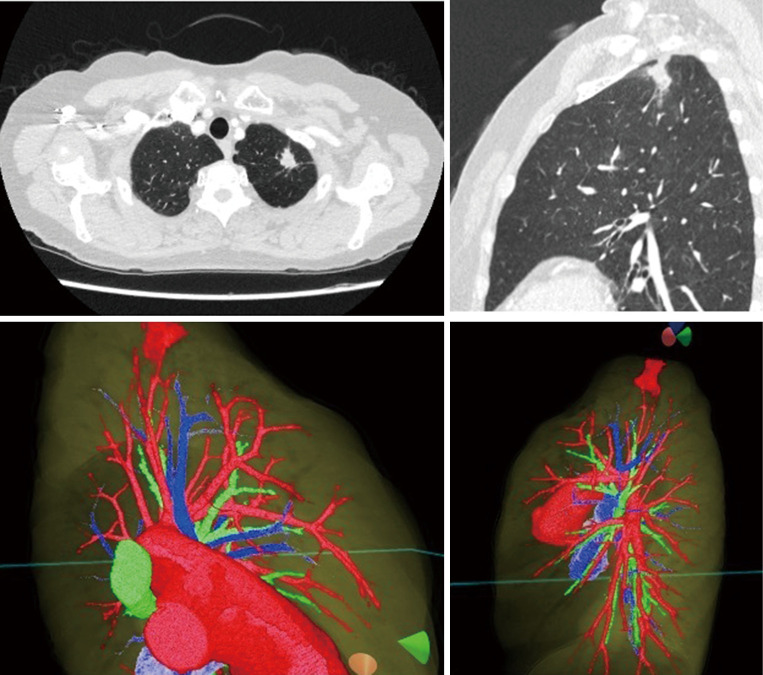
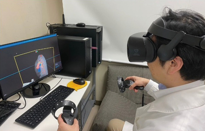
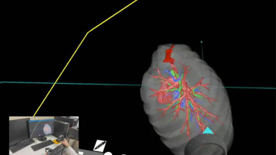
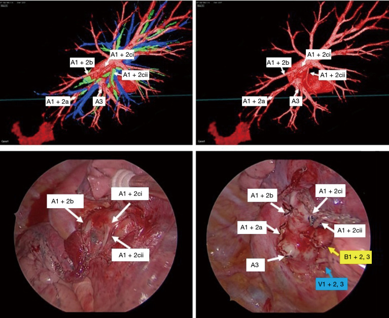
Three-dimensional (3D) polygon data derived from preoperative computed tomography data was loaded into BananaVision software developed at Colorado State University and displayed on an HMD. An interactive 3D reconstruction image was created, in which all the pulmonary structures could be individually imaged. Preoperative resection simulations were performed with patient-individualized reconstructed 3D images.
The 3D anatomic structure of pulmonary vessels and a clear vision into the space between the lesion and adjacent tissues were successfully appreciated during preoperative simulation. Surgeons could easily evaluate the real patient's anatomy in preoperative simulations to improve the accuracy and safety of actual surgery. The VR software and HMD allowed surgeons to visualize and interact with real patient data in true 3D providing a unique perspective.
This initial experience suggests that a VR simulation with HMD facilitated preoperative simulation. Routine imaging modalities combined with VR systems could substantially improve preoperative planning and contribute to the safety and accuracy of anatomic resection.
电视辅助胸腔镜手术(VATS)已成为治疗肺癌的标准方法。然而,其微创性质限制了视野并减少了触觉反馈。这些限制使得外科医生在术前彻底熟悉患者的解剖结构至关重要。我们开发了一种使用头戴式显示器(HMD)的虚拟现实(VR)手术导航系统。本研究的目的是调查这种VR模拟系统在术前规划和术中辅助方面的潜在效用,包括在胸腔镜亚肺叶切除术中提供支持。
将术前计算机断层扫描数据导出的三维(3D)多边形数据加载到科罗拉多州立大学开发的BananaVision软件中,并显示在HMD上。创建了交互式3D重建图像,其中所有肺部结构都可以单独成像。使用患者个体化的重建3D图像进行术前切除模拟。
在术前模拟过程中,成功识别了肺血管的3D解剖结构,并清晰地看到了病变与相邻组织之间的间隙。外科医生可以在术前模拟中轻松评估真实患者的解剖结构,以提高实际手术的准确性和安全性。VR软件和HMD使外科医生能够以真实的3D形式可视化真实患者数据并与之交互,提供了独特的视角。
这一初步经验表明,使用HMD的VR模拟有助于术前模拟。常规成像方式与VR系统相结合可以显著改善术前规划,并有助于解剖性切除的安全性和准确性。