Department of General, Visceral and Transplantation Surgery, University of Heidelberg, Im Neuenheimer Feld 672, 69120, Heidelberg, Germany.
Department of General, Visceral and Thoracic Surgery, University Medical Center Hamburg-Eppendorf, Martinistraße 52, 20246, Hamburg, Germany.
Surg Endosc. 2024 May;38(5):2483-2496. doi: 10.1007/s00464-023-10615-8. Epub 2024 Mar 8.
Evaluation of the benefits of a virtual reality (VR) environment with a head-mounted display (HMD) for decision-making in liver surgery.
Training in liver surgery involves appraising radiologic images and considering the patient's clinical information. Accurate assessment of 2D-tomography images is complex and requires considerable experience, and often the images are divorced from the clinical information. We present a comprehensive and interactive tool for visualizing operation planning data in a VR environment using a head-mounted-display and compare it to 3D visualization and 2D-tomography.
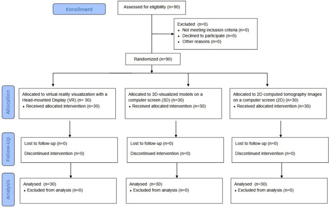
Ninety medical students were randomized into three groups (1:1:1 ratio). All participants analyzed three liver surgery patient cases with increasing difficulty. The cases were analyzed using 2D-tomography data (group "2D"), a 3D visualization on a 2D display (group "3D") or within a VR environment (group "VR"). The VR environment was displayed using the "Oculus Rift ™" HMD technology. Participants answered 11 questions on anatomy, tumor involvement and surgical decision-making and 18 evaluative questions (Likert scale).
Sum of correct answers were significantly higher in the 3D (7.1 ± 1.4, p < 0.001) and VR (7.1 ± 1.4, p < 0.001) groups than the 2D group (5.4 ± 1.4) while there was no difference between 3D and VR (p = 0.987). Times to answer in the 3D (6:44 ± 02:22 min, p < 0.001) and VR (6:24 ± 02:43 min, p < 0.001) groups were significantly faster than the 2D group (09:13 ± 03:10 min) while there was no difference between 3D and VR (p = 0.419). The VR environment was evaluated as most useful for identification of anatomic anomalies, risk and target structures and for the transfer of anatomical and pathological information to the intraoperative situation in the questionnaire.
A VR environment with 3D visualization using a HMD is useful as a surgical training tool to accurately and quickly determine liver anatomy and tumor involvement in surgery.
评估虚拟现实(VR)环境与头戴式显示器(HMD)在肝外科决策中的益处。
肝外科培训涉及评估影像学图像并考虑患者的临床信息。准确评估 2D 断层扫描图像很复杂,需要丰富的经验,并且图像通常与临床信息分离。我们提出了一种全面的交互式工具,用于在 VR 环境中使用头戴式显示器可视化手术计划数据,并将其与 3D 可视化和 2D 断层扫描进行比较。
将 90 名医学生随机分为三组(1:1:1 比例)。所有参与者均分析了三个具有递增难度的肝外科患者病例。这些病例使用 2D 断层扫描数据(组“2D”)、2D 显示器上的 3D 可视化(组“3D”)或 VR 环境(组“VR”)进行分析。VR 环境使用“Oculus Rift ™”HMD 技术显示。参与者回答了 11 个关于解剖结构、肿瘤累及和手术决策的问题以及 18 个评估问题(李克特量表)。
3D(7.1±1.4,p<0.001)和 VR(7.1±1.4,p<0.001)组的正确答案总数明显高于 2D 组(5.4±1.4),而 3D 组和 VR 组之间没有差异(p=0.987)。3D(6:44±02:22 min,p<0.001)和 VR(6:24±02:43 min,p<0.001)组的回答时间明显快于 2D 组(09:13±03:10 min),而 3D 组和 VR 组之间没有差异(p=0.419)。在问卷调查中,VR 环境被评估为识别解剖异常、风险和目标结构以及将解剖和病理信息转移到手术中的最有用工具。
使用 HMD 的 VR 环境与 3D 可视化一起可用作外科培训工具,可准确快速地确定肝脏解剖结构和肿瘤累及情况。