• 文献检索
  • 文档翻译
  • 深度研究
  • 学术资讯
  • Suppr Zotero 插件Zotero 插件
  • 邀请有礼
  • 套餐&价格
  • 历史记录
应用&插件
Suppr Zotero 插件Zotero 插件浏览器插件Mac 客户端Windows 客户端微信小程序
定价
高级版会员购买积分包购买API积分包
服务
文献检索文档翻译深度研究API 文档MCP 服务
关于我们
关于 Suppr公司介绍联系我们用户协议隐私条款
关注我们

Suppr 超能文献

核心技术专利:CN118964589B侵权必究
粤ICP备2023148730 号-1Suppr @ 2026

文献检索

告别复杂PubMed语法,用中文像聊天一样搜索,搜遍4000万医学文献。AI智能推荐,让科研检索更轻松。

立即免费搜索

文件翻译

保留排版,准确专业,支持PDF/Word/PPT等文件格式,支持 12+语言互译。

免费翻译文档

深度研究

AI帮你快速写综述,25分钟生成高质量综述,智能提取关键信息,辅助科研写作。

立即免费体验

基于CT的放射组学分析用于胸腺瘤与胸腺癌的鉴别诊断。

CT-based radiomics analysis for differentiation between thymoma and thymic carcinoma.

作者信息

Ohira Ryosuke, Yanagawa Masahiro, Suzuki Yuki, Hata Akinori, Miyata Tomo, Kikuchi Noriko, Yoshida Yuriko, Yamagata Kazuki, Doi Shuhei, Ninomiya Keisuke, Tomiyama Noriyuki

机构信息

Department of Radiology, Osaka University Graduate School of Medicine, Osaka, Japan.

Department of Artificial Intelligence Diagnostic Radiology, Osaka University Graduate School of Medicine, Osaka, Japan.

出版信息

J Thorac Dis. 2022 May;14(5):1342-1352. doi: 10.21037/jtd-21-1948.

DOI:10.21037/jtd-21-1948
PMID:35693628
原文链接:https://pmc.ncbi.nlm.nih.gov/articles/PMC9186235/
Abstract

BACKGROUND

The purpose of our study was to differentiate between thymoma and thymic carcinoma using a radiomics analysis based on the computed tomography (CT) image features.

METHODS

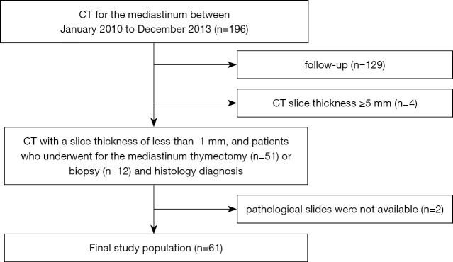
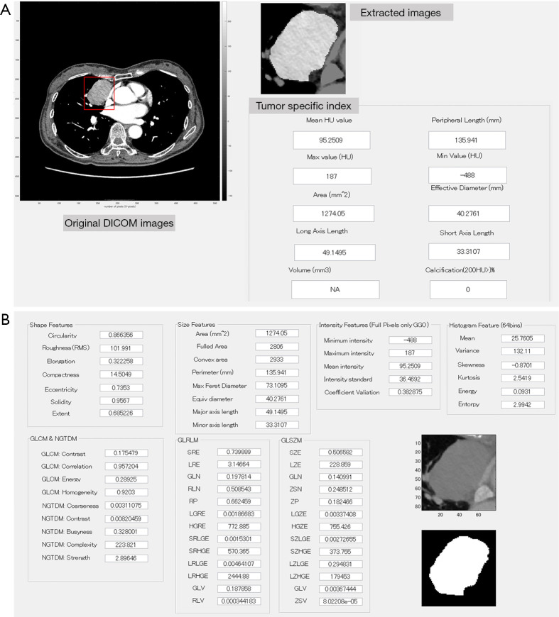
The CT images of 61 patients with thymic epithelial tumors (TETs) who underwent contrast-enhanced CT with slice thickness <1 mm were analyzed. Pathological examination of the surgical specimens revealed thymoma in 45 and thymic carcinoma in 16. Tumor volume and the ratio of major axis to minor axis were calculated using a computer-aided diagnostic software. Sixty-one different radiomics features in the segmented CT images were extracted, then filtered and minimized by least absolute shrinkage and selection operator (LASSO) regression to select the optimal radiomics features for predicting thymic carcinoma. The association between the quantitative values and a diagnosis of thymic carcinoma were analyzed with logistic regression models. Parameters identified as significant in univariate analysis were included in multiple analyses. Receiver-operating characteristic (ROC) curves were assessed to evaluate the diagnostic performance.

RESULTS

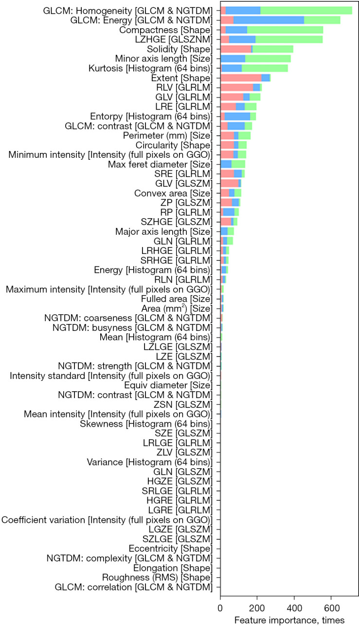
Thymic carcinoma was significantly predominant in men (P=0.001). Optimal radiomics features for predicting thymic carcinoma were as follows: gray-level co-occurrence matrix (GLCM)-homogeneity, GLCM-energy, compactness, large zone high gray-level emphasis (LZHGE), solidity, size of minor axis, and kurtosis. Multiple logistic regression analysis of these features revealed solidity and GLCM-energy as independent indicators associated with thymic carcinoma [odds ratio, 14.7 and 14.3; 95% confidence interval (CI): 1.6-139.0 and 3.0-68.7; and P=0.045 and 0.002, respectively]. Area under the curve (AUC) for diagnosing thymic carcinoma was 0.882 (sensitivity, 81.2%; specificity, 91.1%). Multivariate analysis adjusted for sex similarly revealed two features (solidity and GLCM-energy) as independent indicators associated with thymic carcinoma (odds ratio, 14.6 and 23.9; 95% CI: 2.4-89.2 and 1.9-302.8; P=0.004 and 0.014, respectively). Adjusted AUC for diagnosing thymic carcinoma was 0.921 (95% CI: 0.82-0.97): sensitivity, 62.5% and specificity, 100%.

CONCLUSIONS

Two texture features (GLCM-energy and solidity) were significant predictors of thymic carcinoma.

摘要

背景

我们研究的目的是基于计算机断层扫描(CT)图像特征,通过放射组学分析区分胸腺瘤和胸腺癌。

方法

分析了61例接受层厚<1mm的增强CT检查的胸腺上皮肿瘤(TET)患者的CT图像。手术标本的病理检查显示45例为胸腺瘤,16例为胸腺癌。使用计算机辅助诊断软件计算肿瘤体积和长轴与短轴之比。在分割的CT图像中提取61种不同的放射组学特征,然后通过最小绝对收缩和选择算子(LASSO)回归进行过滤和最小化,以选择预测胸腺癌的最佳放射组学特征。使用逻辑回归模型分析定量值与胸腺癌诊断之间的关联。在单变量分析中确定为显著的参数纳入多变量分析。评估受试者操作特征(ROC)曲线以评估诊断性能。

结果

胸腺癌在男性中显著居多(P=0.001)。预测胸腺癌的最佳放射组学特征如下:灰度共生矩阵(GLCM)-同质性、GLCM-能量、紧密度、大区域高灰度强调(LZHGE)、实性、短轴大小和峰度。对这些特征进行多变量逻辑回归分析,结果显示实性和GLCM-能量是与胸腺癌相关的独立指标[比值比分别为14.7和14.3;95%置信区间(CI):1.6-139.0和3.0-68.7;P分别为0.045和0.002]。诊断胸腺癌的曲线下面积(AUC)为0.882(敏感性为81.2%;特异性为91.1%)。对性别进行校正的多变量分析同样显示两个特征(实性和GLCM-能量)是与胸腺癌相关的独立指标(比值比分别为14.6和23.9;95%CI:2.4-89.2和1.9-302.8;P分别为0.004和0.014)。校正后诊断胸腺癌的AUC为0.921(95%CI:0.82-0.97):敏感性为62.5%,特异性为100%。

结论

两个纹理特征(GLCM-能量和实性)是胸腺癌的重要预测指标。

https://cdn.ncbi.nlm.nih.gov/pmc/blobs/3c6d/9186235/959928ad8a85/jtd-14-05-1342-f3.jpg
https://cdn.ncbi.nlm.nih.gov/pmc/blobs/3c6d/9186235/fea06d0e6b08/jtd-14-05-1342-f1.jpg
https://cdn.ncbi.nlm.nih.gov/pmc/blobs/3c6d/9186235/c43ea1bd9289/jtd-14-05-1342-f2.jpg
https://cdn.ncbi.nlm.nih.gov/pmc/blobs/3c6d/9186235/959928ad8a85/jtd-14-05-1342-f3.jpg
https://cdn.ncbi.nlm.nih.gov/pmc/blobs/3c6d/9186235/fea06d0e6b08/jtd-14-05-1342-f1.jpg
https://cdn.ncbi.nlm.nih.gov/pmc/blobs/3c6d/9186235/c43ea1bd9289/jtd-14-05-1342-f2.jpg
https://cdn.ncbi.nlm.nih.gov/pmc/blobs/3c6d/9186235/959928ad8a85/jtd-14-05-1342-f3.jpg

相似文献

1
CT-based radiomics analysis for differentiation between thymoma and thymic carcinoma.基于CT的放射组学分析用于胸腺瘤与胸腺癌的鉴别诊断。
J Thorac Dis. 2022 May;14(5):1342-1352. doi: 10.21037/jtd-21-1948.
2
Computed tomography radiomic feature analysis of thymic epithelial tumors: Differentiation of thymic epithelial tumors from thymic cysts and prediction of histological subtypes.计算机断层扫描放射组学特征分析胸腺瘤:胸腺瘤与胸囊肿的鉴别及组织学分型预测。
Jpn J Radiol. 2024 Apr;42(4):367-373. doi: 10.1007/s11604-023-01512-0. Epub 2023 Nov 27.
3
CT-Based Radiomics Signatures for Predicting the Risk Categorization of Thymic Epithelial Tumors.基于CT的影像组学特征预测胸腺上皮肿瘤的风险分类
Front Oncol. 2021 Feb 26;11:628534. doi: 10.3389/fonc.2021.628534. eCollection 2021.
4
A radiomics model to predict the invasiveness of thymic epithelial tumors based on contrast‑enhanced computed tomography.基于增强 CT 的影像组学模型预测胸腺瘤侵袭性
Oncol Rep. 2020 Apr;43(4):1256-1266. doi: 10.3892/or.2020.7497. Epub 2020 Feb 11.
5
Usefulness of Three-Dimensional Iodine Mapping Quantified by Dual-Energy CT for Differentiating Thymic Epithelial Tumors.双能CT定量三维碘映射在鉴别胸腺上皮肿瘤中的应用价值
J Clin Med. 2023 Aug 28;12(17):5610. doi: 10.3390/jcm12175610.
6
Optimizing the radiomics-machine-learning model based on non-contrast enhanced CT for the simplified risk categorization of thymic epithelial tumors: A large cohort retrospective study.基于非增强CT优化放射组学-机器学习模型用于胸腺瘤简化风险分类:一项大型队列回顾性研究
Lung Cancer. 2022 Apr;166:150-160. doi: 10.1016/j.lungcan.2022.03.007. Epub 2022 Mar 8.
7
The predictive value of a computed tomography-based radiomics model for the surgical separability of thymic epithelial tumors from the superior vena cava and the left innominate vein.基于计算机断层扫描的影像组学模型对胸腺上皮肿瘤与上腔静脉及左无名静脉手术可分离性的预测价值。
Quant Imaging Med Surg. 2023 Sep 1;13(9):5622-5640. doi: 10.21037/qims-22-1050. Epub 2023 Jul 3.
8
Radiomics Analysis of Multiphasic Computed Tomography Images for Distinguishing High-Risk Thymic Epithelial Tumors From Low-Risk Thymic Epithelial Tumors.多期计算机断层扫描图像的放射组学分析,用于鉴别高危胸腺上皮肿瘤和低危胸腺上皮肿瘤。
J Comput Assist Tomogr. 2023;47(2):220-228. doi: 10.1097/RCT.0000000000001407. Epub 2022 Dec 13.
9
Quantitative CT parameters combined with preoperative systemic inflammatory markers for differentiating risk subgroups of thymic epithelial tumors.定量 CT 参数联合术前全身炎症标志物鉴别胸腺瘤危险亚组。
BMC Cancer. 2023 Nov 27;23(1):1158. doi: 10.1186/s12885-023-11332-0.
10
Contrast-enhanced CT-based radiomics model for differentiating risk subgroups of thymic epithelial tumors.基于增强 CT 的影像组学模型鉴别胸腺瘤的危险亚组。
BMC Med Imaging. 2022 Mar 6;22(1):37. doi: 10.1186/s12880-022-00768-8.

引用本文的文献

1
Using optimized CT type to predict histological classifications of thymic epithelial tumors: a radiomics integrated analysis.利用优化的CT类型预测胸腺上皮肿瘤的组织学分类:一项影像组学综合分析
Insights Imaging. 2025 Mar 22;16(1):67. doi: 10.1186/s13244-025-01933-7.
2
Radiomics in thymic epithelial tumors: a scoping review of current status and advances.胸腺瘤的影像组学:现状与进展的概述性综述
BMC Cancer. 2025 Mar 17;25(1):493. doi: 10.1186/s12885-025-13909-3.
3
Risk stratification of thymic epithelial tumors based on peritumor CT radiomics and semantic features.

本文引用的文献

1
MR imaging of thymomas: a combined radiomics nomogram to predict histologic subtypes.胸腺瘤的 MRI 表现:一种联合放射组学列线图预测组织学亚型。
Eur Radiol. 2021 Jan;31(1):447-457. doi: 10.1007/s00330-020-07074-3. Epub 2020 Jul 22.
2
A radiomics model to predict the invasiveness of thymic epithelial tumors based on contrast‑enhanced computed tomography.基于增强 CT 的影像组学模型预测胸腺瘤侵袭性
Oncol Rep. 2020 Apr;43(4):1256-1266. doi: 10.3892/or.2020.7497. Epub 2020 Feb 11.
3
The Image Biomarker Standardization Initiative: Standardized Quantitative Radiomics for High-Throughput Image-based Phenotyping.
基于肿瘤周围CT影像组学和语义特征的胸腺上皮肿瘤风险分层
Insights Imaging. 2024 Oct 22;15(1):253. doi: 10.1186/s13244-024-01798-2.
4
Preoperative discrimination of invasive and non-invasive breast cancer using machine learning based on automated breast volume scanning (ABVS) radiomics and virtual touch quantification (VTQ).基于自动乳腺容积扫描(ABVS)影像组学和虚拟触诊定量(VTQ),运用机器学习对浸润性和非浸润性乳腺癌进行术前鉴别。
Discov Oncol. 2024 Oct 16;15(1):565. doi: 10.1007/s12672-024-01438-7.
5
The predictive value of a computed tomography-based radiomics model for the surgical separability of thymic epithelial tumors from the superior vena cava and the left innominate vein.基于计算机断层扫描的影像组学模型对胸腺上皮肿瘤与上腔静脉及左无名静脉手术可分离性的预测价值。
Quant Imaging Med Surg. 2023 Sep 1;13(9):5622-5640. doi: 10.21037/qims-22-1050. Epub 2023 Jul 3.
6
Usefulness of Three-Dimensional Iodine Mapping Quantified by Dual-Energy CT for Differentiating Thymic Epithelial Tumors.双能CT定量三维碘映射在鉴别胸腺上皮肿瘤中的应用价值
J Clin Med. 2023 Aug 28;12(17):5610. doi: 10.3390/jcm12175610.
7
Current Treatment Approaches for Thymic Epithelial Tumors.胸腺上皮肿瘤的当前治疗方法
Life (Basel). 2023 May 12;13(5):1170. doi: 10.3390/life13051170.
影像生物标志物标准化倡议:高通量基于影像表型的标准化定量放射组学。
Radiology. 2020 May;295(2):328-338. doi: 10.1148/radiol.2020191145. Epub 2020 Mar 10.
4
Overview of radiomics in breast cancer diagnosis and prognostication.乳腺癌诊断和预后中的放射组学概述。
Breast. 2020 Feb;49:74-80. doi: 10.1016/j.breast.2019.10.018. Epub 2019 Nov 6.
5
CT-based radiomics features analysis for predicting the risk of anterior mediastinal lesions.基于CT的影像组学特征分析用于预测前纵隔病变风险
J Thorac Dis. 2019 May;11(5):1809-1818. doi: 10.21037/jtd.2019.05.32.
6
Radiomics Signatures of Computed Tomography Imaging for Predicting Risk Categorization and Clinical Stage of Thymomas.基于 CT 影像学的影像组学特征预测胸腺瘤风险分级和临床分期
Biomed Res Int. 2019 May 28;2019:3616852. doi: 10.1155/2019/3616852. eCollection 2019.
7
Systemic treatments for thymoma and thymic carcinoma: A systematic review.胸腺瘤和胸腺癌的系统治疗:系统评价。
Lung Cancer. 2018 Dec;126:25-31. doi: 10.1016/j.lungcan.2018.10.018. Epub 2018 Oct 18.
8
Volumetric analysis of the thymic epithelial tumors: correlation of tumor volume with the WHO classification and Masaoka staging.胸腺上皮肿瘤的体积分析:肿瘤体积与世界卫生组织分类及Masaoka分期的相关性
J Thorac Dis. 2018 Oct;10(10):5822-5832. doi: 10.21037/jtd.2018.09.133.
9
Radiomics of pulmonary nodules and lung cancer.肺结节与肺癌的影像组学
Transl Lung Cancer Res. 2017 Feb;6(1):86-91. doi: 10.21037/tlcr.2017.01.04.
10
Volume-based quantification using dual-energy computed tomography in the differentiation of thymic epithelial tumours: an initial experience.基于容积的双能计算机断层扫描在胸腺上皮肿瘤鉴别诊断中的应用:初步经验
Eur Radiol. 2017 May;27(5):1992-2001. doi: 10.1007/s00330-016-4542-9. Epub 2016 Aug 23.