Biochemistry and Biophysics Center, National Heart Lung and Blood Institute, National Institutes of Health, Bethesda, MD.
Department of Cellular Neurobiology, Graduate School of Medicine, The University of Tokyo, Tokyo, Japan.
J Gen Physiol. 2021 Apr 5;153(4). doi: 10.1085/jgp.202012814.
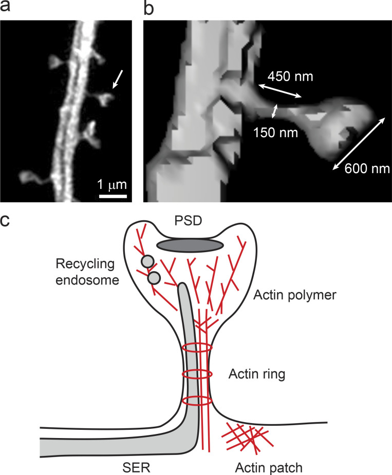
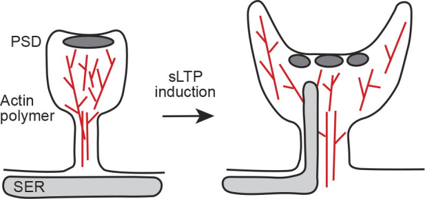
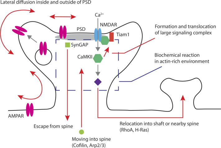
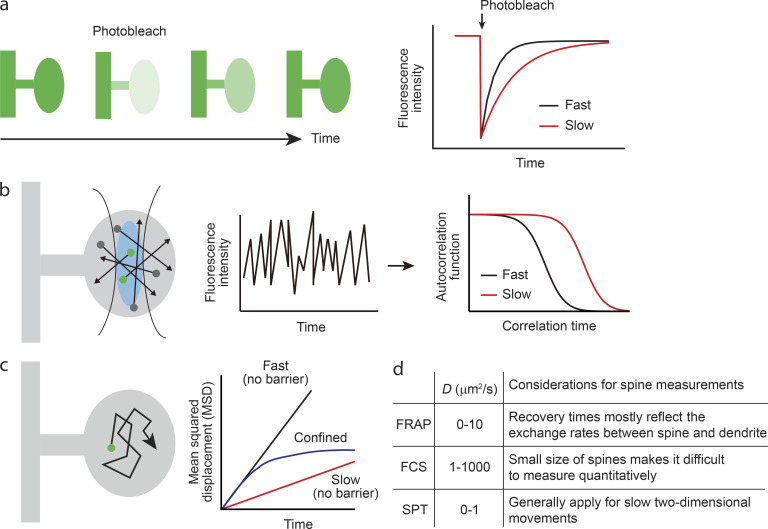
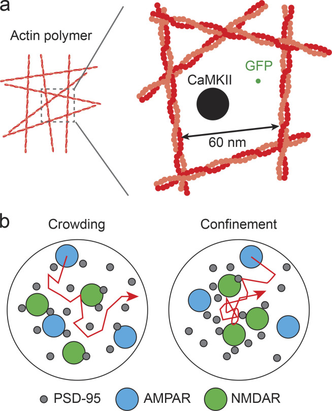
Spines are tiny nanoscale protrusions from dendrites of neurons. In the cortex and hippocampus, most of the excitatory postsynaptic sites reside in spines. The bulbous spine head is connected to the dendritic shaft by a thin membranous neck. Because the neck is narrow, spine heads are thought to function as biochemically independent signaling compartments. Thus, dynamic changes in the composition, distribution, mobility, conformations, and signaling properties of molecules contained within spines can account for much of the molecular basis of postsynaptic function and regulation. A major factor in controlling these changes is the diffusional properties of proteins within this small compartment. Advances in measurement techniques using fluorescence microscopy now make it possible to measure molecular diffusion within single dendritic spines directly. Here, we review the regulatory mechanisms of diffusion in spines by local intra-spine architecture and discuss their implications for neuronal signaling and synaptic plasticity.
棘突是神经元树突上的微小纳米级突起。在大脑皮层和海马体中,大多数兴奋性突触后位点都位于棘突中。球状棘突头部通过细的膜状颈部与树突干相连。由于颈部狭窄,棘突头部被认为是具有生化独立性的信号隔室。因此,包含在棘突内的分子的组成、分布、流动性、构象和信号转导特性的动态变化可以解释突触后功能和调节的大部分分子基础。控制这些变化的一个主要因素是这个小隔室内蛋白质的扩散特性。使用荧光显微镜的测量技术的进步现在使得可以直接测量单个树突棘突内的分子扩散。在这里,我们通过局部棘突内结构来综述扩散的调节机制,并讨论其对神经元信号转导和突触可塑性的影响。