Yip Mighten C, Gonzalez Mercedes M, Valenta Christopher R, Rowan Matthew J M, Forest Craig R
Georgia Institute of Technology, George W. Woodruff School of Mechanical Engineering, Atlanta, 30332, USA.
Georgia Tech Research Institute, Atlanta, 30332, USA.
Sci Rep. 2021 Mar 16;11(1):6065. doi: 10.1038/s41598-021-85695-4.
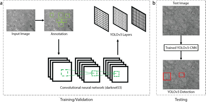
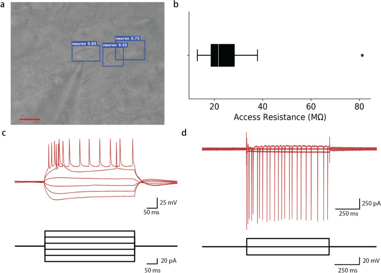
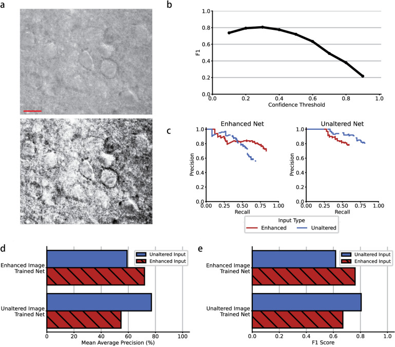
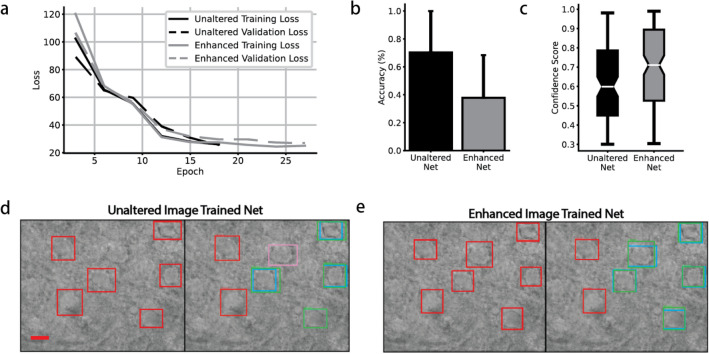
A common electrophysiology technique used in neuroscience is patch clamp: a method in which a glass pipette electrode facilitates single cell electrical recordings from neurons. Typically, patch clamp is done manually in which an electrophysiologist views a brain slice under a microscope, visually selects a neuron to patch, and moves the pipette into close proximity to the cell to break through and seal its membrane. While recent advances in the field of patch clamping have enabled partial automation, the task of detecting a healthy neuronal soma in acute brain tissue slices is still a critical step that is commonly done manually, often presenting challenges for novices in electrophysiology. To overcome this obstacle and progress towards full automation of patch clamp, we combined the differential interference microscopy optical technique with an object detection-based convolutional neural network (CNN) to detect healthy neurons in acute slice. Utilizing the YOLOv3 convolutional neural network architecture, we achieved a 98% reduction in training times to 18 min, compared to previously published attempts. We also compared networks trained on unaltered and enhanced images, achieving up to 77% and 72% mean average precision, respectively. This novel, deep learning-based method accomplishes automated neuronal detection in brain slice at 18 frames per second with a small data set of 1138 annotated neurons, rapid training time, and high precision. Lastly, we verified the health of the identified neurons with a patch clamp experiment where the average access resistance was 29.25 M[Formula: see text] (n = 9). The addition of this technology during live-cell imaging for patch clamp experiments can not only improve manual patch clamping by reducing the neuroscience expertise required to select healthy cells, but also help achieve full automation of patch clamping by nominating cells without human assistance.
一种使用玻璃微吸管电极对神经元进行单细胞电记录的方法。通常,膜片钳是手动操作的,电生理学家在显微镜下观察脑切片,目视选择要膜片钳制的神经元,然后将微吸管移至靠近细胞的位置,以穿透并密封其细胞膜。虽然膜片钳领域的最新进展已实现了部分自动化,但在急性脑组织切片中检测健康神经元胞体的任务仍然是一个关键步骤,通常是手动完成,这对电生理新手来说往往具有挑战性。为了克服这一障碍并朝着膜片钳的完全自动化迈进,我们将微分干涉显微镜光学技术与基于目标检测的卷积神经网络(CNN)相结合,以检测急性切片中的健康神经元。利用YOLOv3卷积神经网络架构,与之前发表的尝试相比,我们将训练时间减少了98%,至18分钟。我们还比较了在未改变和增强图像上训练的网络,平均精度分别达到了77%和72%。这种基于深度学习的新方法以每秒18帧的速度,利用1138个带注释神经元的小数据集、快速的训练时间和高精度,完成了脑切片中神经元的自动检测。最后,我们通过膜片钳实验验证了所识别神经元的健康状况,其中平均接入电阻为29.25 MΩ(n = 9)。在膜片钳实验的活细胞成像过程中添加这项技术,不仅可以通过减少选择健康细胞所需的神经科学专业知识来改进手动膜片钳制,还可以通过在无人工协助的情况下指定细胞来帮助实现膜片钳制的完全自动化。