• 文献检索
  • 文档翻译
  • 深度研究
  • 学术资讯
  • Suppr Zotero 插件Zotero 插件
  • 邀请有礼
  • 套餐&价格
  • 历史记录
应用&插件
Suppr Zotero 插件Zotero 插件浏览器插件Mac 客户端Windows 客户端微信小程序
定价
高级版会员购买积分包购买API积分包
服务
文献检索文档翻译深度研究API 文档MCP 服务
关于我们
关于 Suppr公司介绍联系我们用户协议隐私条款
关注我们

Suppr 超能文献

核心技术专利:CN118964589B侵权必究
粤ICP备2023148730 号-1Suppr @ 2026

文献检索

告别复杂PubMed语法,用中文像聊天一样搜索,搜遍4000万医学文献。AI智能推荐,让科研检索更轻松。

立即免费搜索

文件翻译

保留排版,准确专业,支持PDF/Word/PPT等文件格式,支持 12+语言互译。

免费翻译文档

深度研究

AI帮你快速写综述,25分钟生成高质量综述,智能提取关键信息,辅助科研写作。

立即免费体验

无参分子超结构在单分子定位显微镜中的定量。

Parameter-free molecular super-structures quantification in single-molecule localization microscopy.

机构信息

Medical Research Council Human Genetics Unit, Institute of Genetics and Molecular Medicine, University of Edinburgh, Edinburgh, UK.

Scottish Universities Physics Alliance, School of Physics and Astronomy, University of Edinburgh, Edinburgh, UK.

出版信息

J Cell Biol. 2021 May 3;220(5). doi: 10.1083/jcb.202010003.

DOI:10.1083/jcb.202010003
PMID:33734291
原文链接:https://pmc.ncbi.nlm.nih.gov/articles/PMC7980255/
Abstract

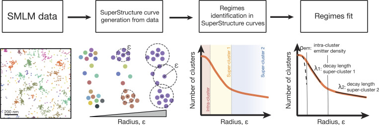
Understanding biological function requires the identification and characterization of complex patterns of molecules. Single-molecule localization microscopy (SMLM) can quantitatively measure molecular components and interactions at resolutions far beyond the diffraction limit, but this information is only useful if these patterns can be quantified and interpreted. We provide a new approach for the analysis of SMLM data that develops the concept of structures and super-structures formed by interconnected elements, such as smaller protein clusters. Using a formal framework and a parameter-free algorithm, (super-)structures formed from smaller components are found to be abundant in classes of nuclear proteins, such as heterogeneous nuclear ribonucleoprotein particles (hnRNPs), but are absent from ceramides located in the plasma membrane. We suggest that mesoscopic structures formed by interconnected protein clusters are common within the nucleus and have an important role in the organization and function of the genome. Our algorithm, SuperStructure, can be used to analyze and explore complex SMLM data and extract functionally relevant information.

摘要

理解生物功能需要识别和描述复杂的分子模式。单分子定位显微镜(SMLM)可以在远远超过衍射极限的分辨率下定量测量分子成分和相互作用,但只有当这些模式可以被定量和解释时,这些信息才有用。我们提供了一种用于分析 SMLM 数据的新方法,该方法发展了由相互连接的元素(如较小的蛋白质簇)形成的结构和超结构的概念。使用正式框架和无参数算法,发现由较小成分形成的(超)结构在核蛋白(如异质核核糖核蛋白颗粒(hnRNPs))等类别中非常丰富,但在位于质膜中的神经酰胺中不存在。我们认为,由相互连接的蛋白质簇形成的介观结构在核内很常见,在基因组的组织和功能中具有重要作用。我们的算法 SuperStructure 可用于分析和探索复杂的 SMLM 数据并提取功能相关信息。

https://cdn.ncbi.nlm.nih.gov/pmc/blobs/19a9/7980255/8fd8e87c2332/JCB_202010003_FigS5.jpg
https://cdn.ncbi.nlm.nih.gov/pmc/blobs/19a9/7980255/2c6dfb585e12/JCB_202010003_Fig1.jpg
https://cdn.ncbi.nlm.nih.gov/pmc/blobs/19a9/7980255/8f2538846462/JCB_202010003_FigS1.jpg
https://cdn.ncbi.nlm.nih.gov/pmc/blobs/19a9/7980255/411c46654727/JCB_202010003_Fig2.jpg
https://cdn.ncbi.nlm.nih.gov/pmc/blobs/19a9/7980255/7046ef7f6356/JCB_202010003_FigS2.jpg
https://cdn.ncbi.nlm.nih.gov/pmc/blobs/19a9/7980255/c32af186424c/JCB_202010003_Fig3.jpg
https://cdn.ncbi.nlm.nih.gov/pmc/blobs/19a9/7980255/d7fdfe1fa06a/JCB_202010003_FigS3.jpg
https://cdn.ncbi.nlm.nih.gov/pmc/blobs/19a9/7980255/35064b2d544f/JCB_202010003_Fig4.jpg
https://cdn.ncbi.nlm.nih.gov/pmc/blobs/19a9/7980255/be69b60135f8/JCB_202010003_FigS4.jpg
https://cdn.ncbi.nlm.nih.gov/pmc/blobs/19a9/7980255/8fd8e87c2332/JCB_202010003_FigS5.jpg

相似文献

1
Parameter-free molecular super-structures quantification in single-molecule localization microscopy.无参分子超结构在单分子定位显微镜中的定量。
J Cell Biol. 2021 May 3;220(5). doi: 10.1083/jcb.202010003.
2
DeepCEL0 for 2D single-molecule localization in fluorescence microscopy.DeepCEL0 用于荧光显微镜 2D 单分子定位。
Bioinformatics. 2022 Feb 7;38(5):1411-1419. doi: 10.1093/bioinformatics/btab808.
3
Chemistry of Photosensitive Fluorophores for Single-Molecule Localization Microscopy.用于单分子定位显微镜的光敏荧光团化学
ACS Chem Biol. 2019 Jun 21;14(6):1077-1090. doi: 10.1021/acschembio.9b00197. Epub 2019 May 13.
4
Accelerating single molecule localization microscopy through parallel processing on a high-performance computing cluster.通过在高性能计算集群上进行并行处理来加速单分子定位显微镜技术。
J Microsc. 2019 Feb;273(2):148-160. doi: 10.1111/jmi.12772. Epub 2018 Dec 3.
5
Conventional BODIPY Conjugates for Live-Cell Super-Resolution Microscopy and Single-Molecule Tracking.用于活细胞超分辨率显微镜和单分子追踪的传统硼二吡咯亚甲基缀合物
J Vis Exp. 2020 Jun 8(160). doi: 10.3791/60950.
6
Correlative super-resolution microscopy with deep UV reactivation.深紫外光再激活相关超分辨显微镜。
J Microsc. 2024 Nov;296(2):133-138. doi: 10.1111/jmi.13258. Epub 2024 Jan 3.
7
Quantitative Single-Molecule Localization Microscopy (qSMLM) of Membrane Proteins Based on Kinetic Analysis of Fluorophore Blinking Cycles.基于荧光团闪烁循环动力学分析的膜蛋白定量单分子定位显微镜技术
Methods Mol Biol. 2017;1663:115-126. doi: 10.1007/978-1-4939-7265-4_10.
8
Toward Absolute Molecular Numbers in DNA-PAINT.实现 DNA-PAINT 中的绝对分子数。
Nano Lett. 2019 Nov 13;19(11):8182-8190. doi: 10.1021/acs.nanolett.9b03546. Epub 2019 Oct 9.
9
Photochemical Mechanisms of Fluorophores Employed in Single-Molecule Localization Microscopy.用于单分子定位显微镜的荧光团的光化学机制。
Angew Chem Int Ed Engl. 2023 Jan 2;62(1):e202204745. doi: 10.1002/anie.202204745. Epub 2022 Nov 17.
10
Quantitative super-resolution single molecule microscopy dataset of YFP-tagged growth factor receptors.YFP 标记的生长因子受体的定量超分辨率单分子显微镜数据集。
Gigascience. 2018 Mar 1;7(3):1-10. doi: 10.1093/gigascience/giy002.

引用本文的文献

1
Cryosectioning-enhanced super-resolution microscopy for single-protein imaging across cells and tissues.用于跨细胞和组织的单蛋白成像的冷冻切片增强超分辨率显微镜技术。
Proc Natl Acad Sci U S A. 2025 Aug 12;122(32):e2504578122. doi: 10.1073/pnas.2504578122. Epub 2025 Aug 7.
2
Chromatin-associated α-satellite RNA maintains chromosome stability by reestablishing SAF-A in the mitotic cell cycle.与染色质相关的α-卫星RNA通过在有丝分裂细胞周期中重新建立SAF-A来维持染色体稳定性。
Nucleic Acids Res. 2025 Apr 10;53(7). doi: 10.1093/nar/gkaf294.
3
Quality control maps: Real-time quantitative control of single-molecule localization microscopy data.

本文引用的文献

1
Super-resolution in situ analysis of active ribosomal DNA chromatin organization in the nucleolus.核仁中活性核糖体 DNA 染色质组织的超高分辨率原位分析。
Sci Rep. 2020 May 4;10(1):7462. doi: 10.1038/s41598-020-64589-x.
2
Repetitive RNAs as Regulators of Chromatin-Associated Subcompartment Formation by Phase Separation.重复 RNA 作为相分离调控染色质相关亚区形成的分子。
J Mol Biol. 2020 Jul 10;432(15):4270-4286. doi: 10.1016/j.jmb.2020.04.015. Epub 2020 Apr 19.
3
3D ATAC-PALM: super-resolution imaging of the accessible genome.
质量控制图:单分子定位显微镜数据的实时定量控制
Biophys J. 2025 Apr 1;124(7):1132-1145. doi: 10.1016/j.bpj.2025.02.018. Epub 2025 Feb 25.
4
Edge roughness analysis in nanoscale for single-molecule localization microscopy images.单分子定位显微镜图像的纳米级边缘粗糙度分析
Nanophotonics. 2024 Jan 4;13(2):195-207. doi: 10.1515/nanoph-2023-0709. eCollection 2024 Jan.
5
Subcellular Drug Distribution: Exploring Organelle-Specific Characteristics for Enhanced Therapeutic Efficacy.亚细胞药物分布:探索细胞器特异性特征以提高治疗效果。
Pharmaceutics. 2024 Sep 4;16(9):1167. doi: 10.3390/pharmaceutics16091167.
6
Cryosectioning-enhanced super-resolution microscopy for single-protein imaging across cells and tissues.用于跨细胞和组织的单蛋白成像的冷冻切片增强超分辨率显微镜技术。
bioRxiv. 2025 Feb 13:2024.02.05.576943. doi: 10.1101/2024.02.05.576943.
7
Restrictor synergizes with Symplekin and PNUTS to terminate extragenic transcription.限制因子与 Symplekin 和 PNUTS 协同作用,终止基因外转录。
Genes Dev. 2023 Dec 26;37(21-24):1017-1040. doi: 10.1101/gad.351057.123.
8
Recent development of computational cluster analysis methods for single-molecule localization microscopy images.单分子定位显微镜图像计算聚类分析方法的最新进展。
Comput Struct Biotechnol J. 2023 Jan 9;21:879-888. doi: 10.1016/j.csbj.2023.01.006. eCollection 2023.
9
Unbiased choice of global clustering parameters for single-molecule localization microscopy.无偏全局聚类参数选择用于单分子定位显微镜。
Sci Rep. 2022 Dec 29;12(1):22561. doi: 10.1038/s41598-022-27074-1.
10
Efficient Cross-Correlation Filtering of One- and Two-Color Single Molecule Localization Microscopy Data.单分子定位显微镜数据的高效互相关滤波:单色和双色情况
Front Bioinform. 2021 Nov 4;1:739769. doi: 10.3389/fbinf.2021.739769. eCollection 2021.
3D ATAC-PALM:可及基因组的超高分辨率成像。
Nat Methods. 2020 Apr;17(4):430-436. doi: 10.1038/s41592-020-0775-2. Epub 2020 Mar 16.
4
Machine learning for cluster analysis of localization microscopy data.基于机器学习的定位显微镜数据分析聚类。
Nat Commun. 2020 Mar 20;11(1):1493. doi: 10.1038/s41467-020-15293-x.
5
Chromatin Is Frequently Unknotted at the Megabase Scale.染色质在兆碱基尺度上常呈解结状态。
Biophys J. 2020 May 5;118(9):2268-2279. doi: 10.1016/j.bpj.2019.11.002. Epub 2019 Nov 13.
6
Chromosome dynamics near the sol-gel phase transition dictate the timing of remote genomic interactions.临近溶胶-凝胶转变时的染色体动力学决定了远程基因组相互作用的时间。
Nat Commun. 2019 Jun 24;10(1):2771. doi: 10.1038/s41467-019-10628-9.
7
Evidence for DNA-mediated nuclear compartmentalization distinct from phase separation.有证据表明 DNA 介导的核区室化不同于相分离。
Elife. 2019 May 7;8:e47098. doi: 10.7554/eLife.47098.
8
Role of nuclear RNA in regulating chromatin structure and transcription.核 RNA 在调节染色质结构和转录中的作用。
Curr Opin Cell Biol. 2019 Jun;58:120-125. doi: 10.1016/j.ceb.2019.03.007. Epub 2019 Apr 19.
9
Walking along chromosomes with super-resolution imaging, contact maps, and integrative modeling.用超高分辨率成像、接触图谱和整合建模沿着染色体行走。
PLoS Genet. 2018 Dec 26;14(12):e1007872. doi: 10.1371/journal.pgen.1007872. eCollection 2018 Dec.
10
Super-resolution chromatin tracing reveals domains and cooperative interactions in single cells.超分辨率染色质追踪揭示了单细胞中的域和协同相互作用。
Science. 2018 Oct 26;362(6413). doi: 10.1126/science.aau1783.