Trebeschi Stefano, Bodalal Zuhir, Boellaard Thierry N, Tareco Bucho Teresa M, Drago Silvia G, Kurilova Ieva, Calin-Vainak Adriana M, Delli Pizzi Andrea, Muller Mirte, Hummelink Karlijn, Hartemink Koen J, Nguyen-Kim Thi Dan Linh, Smit Egbert F, Aerts Hugo J W L, Beets-Tan Regina G H
Department of Radiology, Netherlands Cancer Institute - Antoni vanLeeuwenhoek Hospital, Amsterdam, Netherlands.
GROW School for Oncology and Developmental Biology, Maastricht, Netherlands.
Front Oncol. 2021 Mar 2;11:609054. doi: 10.3389/fonc.2021.609054. eCollection 2021.
Checkpoint inhibitors provided sustained clinical benefit to metastatic lung cancer patients. Nonetheless, prognostic markers in metastatic settings are still under research. Imaging offers distinctive advantages, providing whole-body information non-invasively, while routinely available in most clinics. We hypothesized that more prognostic information can be extracted by employing artificial intelligence (AI) for treatment monitoring, superior to 2D tumor growth criteria.
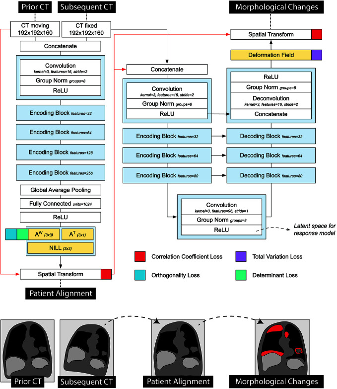
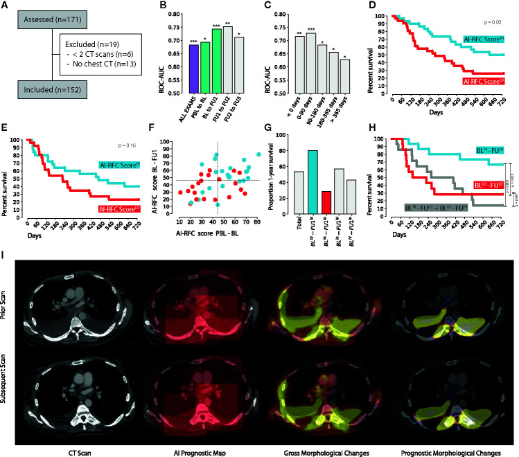
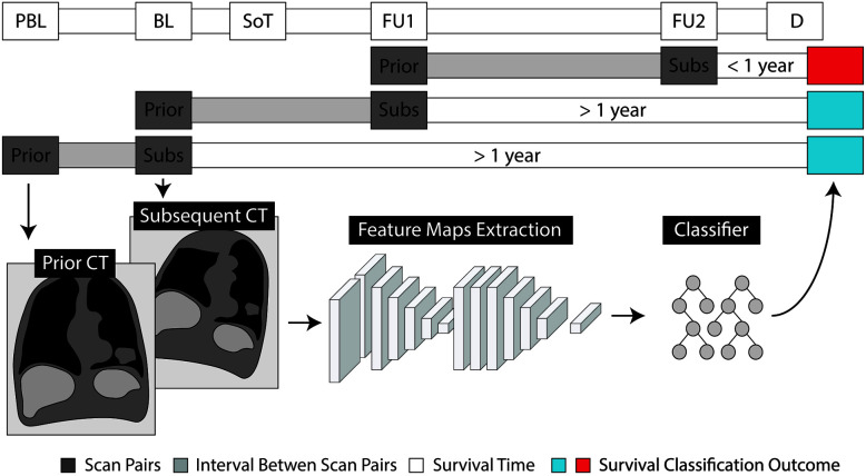
A cohort of 152 stage-IV non-small-cell lung cancer patients (NSCLC) (73 discovery, 79 test, 903CTs), who received nivolumab were retrospectively collected. We trained a neural network to identify morphological changes on chest CT acquired during patients' follow-ups. A classifier was employed to link imaging features learned by the network with overall survival.
Our results showed significant performance in the independent test set to predict 1-year overall survival from the date of image acquisition, with an average area under the curve (AUC) of 0.69 ( 0.01), up to AUC 0.75 ( < 0.01) in the first 3 to 5 months of treatment, and 0.67 AUC ( = 0.01) for durable clinical benefit (6 months progression-free survival). We found the AI-derived survival score to be independent of clinical, radiological, PDL1, and histopathological factors. Visual analysis of AI-generated prognostic heatmaps revealed relative prognostic importance of morphological nodal changes in the mediastinum, supraclavicular, and hilar regions, lung and bone metastases, as well as pleural effusions, atelectasis, and consolidations.
Our results demonstrate that deep learning can quantify tumor- and non-tumor-related morphological changes important for prognostication on serial imaging. Further investigation should focus on the implementation of this technique beyond thoracic imaging.
检查点抑制剂为转移性肺癌患者带来了持续的临床益处。尽管如此,转移性环境中的预后标志物仍在研究中。影像学具有独特的优势,可无创提供全身信息,且在大多数诊所中常规可用。我们假设通过使用人工智能(AI)进行治疗监测可以提取更多的预后信息,优于二维肿瘤生长标准。
回顾性收集了152例接受纳武单抗治疗的IV期非小细胞肺癌(NSCLC)患者(73例用于发现,79例用于测试,903次CT扫描)。我们训练了一个神经网络来识别患者随访期间获得的胸部CT上的形态变化。使用分类器将网络学习到的影像特征与总生存期联系起来。
我们的结果显示,在独立测试集中,从图像采集日期预测1年总生存期具有显著性能,曲线下平均面积(AUC)为0.69(±0.01),在治疗的前3至5个月中AUC高达0.75(P<0.01),对于持久临床获益(6个月无进展生存期)AUC为0.67(P = 0.01)。我们发现人工智能得出的生存评分独立于临床、放射学、PDL1和组织病理学因素。对人工智能生成的预后热图的视觉分析揭示了纵隔、锁骨上和肺门区域、肺和骨转移以及胸腔积液、肺不张和实变中形态学淋巴结变化的相对预后重要性。
我们的结果表明,深度学习可以量化对连续成像预后重要的肿瘤和非肿瘤相关形态学变化。进一步的研究应集中在该技术在胸部成像以外的应用。