• 文献检索
  • 文档翻译
  • 深度研究
  • 学术资讯
  • Suppr Zotero 插件Zotero 插件
  • 邀请有礼
  • 套餐&价格
  • 历史记录
应用&插件
Suppr Zotero 插件Zotero 插件浏览器插件Mac 客户端Windows 客户端微信小程序
定价
高级版会员购买积分包购买API积分包
服务
文献检索文档翻译深度研究API 文档MCP 服务
关于我们
关于 Suppr公司介绍联系我们用户协议隐私条款
关注我们

Suppr 超能文献

核心技术专利:CN118964589B侵权必究
粤ICP备2023148730 号-1Suppr @ 2026

文献检索

告别复杂PubMed语法,用中文像聊天一样搜索,搜遍4000万医学文献。AI智能推荐,让科研检索更轻松。

立即免费搜索

文件翻译

保留排版,准确专业,支持PDF/Word/PPT等文件格式,支持 12+语言互译。

免费翻译文档

深度研究

AI帮你快速写综述,25分钟生成高质量综述,智能提取关键信息,辅助科研写作。

立即免费体验

相似文献

1
Impact of brain shift on neural pathways in deep brain stimulation: a preliminary analysis via multi-physics finite element models.脑移位对脑深部刺激神经通路的影响:基于多物理有限元模型的初步分析。
J Neural Eng. 2021 Apr 6;18(5). doi: 10.1088/1741-2552/abf066.
2
Accounting for Deformation in Deep Brain Stimulation Surgery With Models: Comparison to Interventional Magnetic Resonance Imaging.利用模型对深部脑刺激手术中的变形进行定量分析:与介入磁共振成像的比较。
IEEE Trans Biomed Eng. 2020 Oct;67(10):2934-2944. doi: 10.1109/TBME.2020.2974102. Epub 2020 Feb 14.
3
Deep brain stimulation induced normalization of the human functional connectome in Parkinson's disease.深部脑刺激可使帕金森病患者的功能连接组正常化。
Brain. 2019 Oct 1;142(10):3129-3143. doi: 10.1093/brain/awz239.
4
Clinical deep brain stimulation strategies for orientation-selective pathway activation.临床深部脑刺激策略用于定向选择途径的激活。
J Neural Eng. 2018 Oct;15(5):056029. doi: 10.1088/1741-2552/aad978. Epub 2018 Aug 10.
5
Speech changes induced by deep brain stimulation of the subthalamic nucleus in Parkinson disease: involvement of the dentatorubrothalamic tract.深部脑刺激丘脑底核诱导的帕金森病语音改变:齿状红核束的参与。
J Neurosurg. 2017 Jun;126(6):2017-2027. doi: 10.3171/2016.5.JNS16243. Epub 2016 Sep 9.
6
Impact of brain shift on subcallosal cingulate deep brain stimulation.脑移位对扣带回下深部脑刺激的影响。
Brain Stimul. 2018 Mar-Apr;11(2):445-453. doi: 10.1016/j.brs.2017.12.001. Epub 2017 Dec 6.
7
Model-Based Image Updating for Brain Shift in Deep Brain Stimulation Electrode Placement Surgery.基于模型的脑移位图像更新在深部脑刺激电极植入术中的应用。
IEEE Trans Biomed Eng. 2020 Dec;67(12):3542-3552. doi: 10.1109/TBME.2020.2990669. Epub 2020 Nov 19.
8
Targeting Neuronal Fiber Tracts for Deep Brain Stimulation Therapy Using Interactive, Patient-Specific Models.使用交互式、患者特异性模型靶向神经元纤维束进行深部脑刺激治疗
J Vis Exp. 2018 Aug 12(138):57292. doi: 10.3791/57292.
9
Computational modeling of an endovascular approach to deep brain stimulation.深部脑刺激血管内治疗方法的计算建模
J Neural Eng. 2014 Apr;11(2):026011. doi: 10.1088/1741-2560/11/2/026011. Epub 2014 Mar 10.
10
Model-based image updating in deep brain stimulation with assimilation of deep brain sparse data.基于模型的深部脑刺激中的图像更新,结合深部脑稀疏数据的同化。
Med Phys. 2023 Dec;50(12):7904-7920. doi: 10.1002/mp.16578. Epub 2023 Jul 7.

引用本文的文献

1
Translating high-precision mixed reality navigation from lab to operating room: design and clinical evaluation.将高精度混合现实导航从实验室转化至手术室:设计与临床评估
BMC Surg. 2025 Aug 1;25(1):331. doi: 10.1186/s12893-025-03096-0.
2
Advances in the intraoperative delineation of malignant glioma margin.恶性胶质瘤边缘术中划定的进展
Front Oncol. 2023 Jan 26;13:1114450. doi: 10.3389/fonc.2023.1114450. eCollection 2023.
3
Automatic framework for patient-specific modelling of tumour resection-induced brain shift.用于肿瘤切除引起的脑移位的患者特异性建模的自动框架。
Comput Biol Med. 2022 Apr;143:105271. doi: 10.1016/j.compbiomed.2022.105271. Epub 2022 Jan 30.

本文引用的文献

1
Postoperative Lead Movement after Deep Brain Stimulation Surgery and the Change of Stimulation Volume.术后深部脑刺激手术后的导联移动和刺激量的变化。
Stereotact Funct Neurosurg. 2021;99(3):221-229. doi: 10.1159/000511406. Epub 2020 Dec 16.
2
Orientation-selective and directional deep brain stimulation in swine assessed by functional MRI at 3T.3T 功能磁共振成像评估猪的取向选择性和方向选择性深部脑刺激。
Neuroimage. 2021 Jan 1;224:117357. doi: 10.1016/j.neuroimage.2020.117357. Epub 2020 Sep 9.
3
Normative vs. patient-specific brain connectivity in deep brain stimulation.规范与个体化脑连接在脑深部刺激中的比较。
Neuroimage. 2021 Jan 1;224:117307. doi: 10.1016/j.neuroimage.2020.117307. Epub 2020 Aug 28.
4
Accounting for intraoperative brain shift ascribable to cavity collapse during intracranial tumor resection.颅内肿瘤切除术中因腔隙塌陷导致的术中脑移位的计算。
J Med Imaging (Bellingham). 2020 May;7(3):031506. doi: 10.1117/1.JMI.7.3.031506. Epub 2020 Jun 22.
5
Accounting for Deformation in Deep Brain Stimulation Surgery With Models: Comparison to Interventional Magnetic Resonance Imaging.利用模型对深部脑刺激手术中的变形进行定量分析:与介入磁共振成像的比较。
IEEE Trans Biomed Eng. 2020 Oct;67(10):2934-2944. doi: 10.1109/TBME.2020.2974102. Epub 2020 Feb 14.
6
Evaluation of methodologies for computing the deep brain stimulation volume of tissue activated.组织激活的深部脑刺激体积计算方法的评估。
J Neural Eng. 2019 Oct 29;16(6):066024. doi: 10.1088/1741-2552/ab3c95.
7
Subthalamic neuromodulation improves short-term motor learning in Parkinson's disease.丘脑底核电刺激改善帕金森病的短期运动学习。
Brain. 2019 Aug 1;142(8):2198-2206. doi: 10.1093/brain/awz152.
8
Efficacy and Safety of Deep Brain Stimulation in the Treatment of Parkinson's Disease: A Systematic Review and Meta-analysis of Randomized Controlled Trials.深部脑刺激治疗帕金森病的疗效与安全性:随机对照试验的系统评价与Meta分析
Cureus. 2018 Oct 22;10(10):e3474. doi: 10.7759/cureus.3474.
9
Accuracy of Intraoperative Computed Tomography in Deep Brain Stimulation-A Prospective Noninferiority Study.术中计算机断层扫描在脑深部刺激中的准确性:一项前瞻性非劣效性研究。
Neuromodulation. 2019 Jun;22(4):472-477. doi: 10.1111/ner.12918. Epub 2019 Jan 10.
10
Thalamic and ventricular volumes predict motor response to deep brain stimulation for Parkinson's disease.丘脑和脑室体积可预测帕金森病深部脑刺激的运动反应。
Parkinsonism Relat Disord. 2019 Apr;61:64-69. doi: 10.1016/j.parkreldis.2018.11.026. Epub 2018 Nov 28.

脑移位对脑深部刺激神经通路的影响:基于多物理有限元模型的初步分析。

Impact of brain shift on neural pathways in deep brain stimulation: a preliminary analysis via multi-physics finite element models.

机构信息

Department of Biomedical Engineering, Vanderbilt University, Nashville, TN, United States of America.

Vanderbilt Institute for Surgery and Engineering, Vanderbilt University, Nashville, TN, United States of America.

出版信息

J Neural Eng. 2021 Apr 6;18(5). doi: 10.1088/1741-2552/abf066.

DOI:10.1088/1741-2552/abf066
PMID:33740780
原文链接:https://pmc.ncbi.nlm.nih.gov/articles/PMC9476060/
Abstract

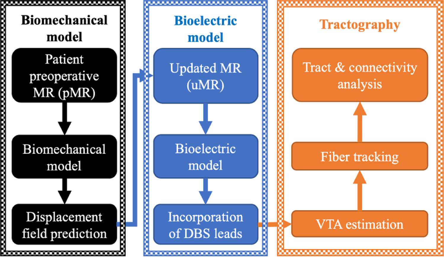
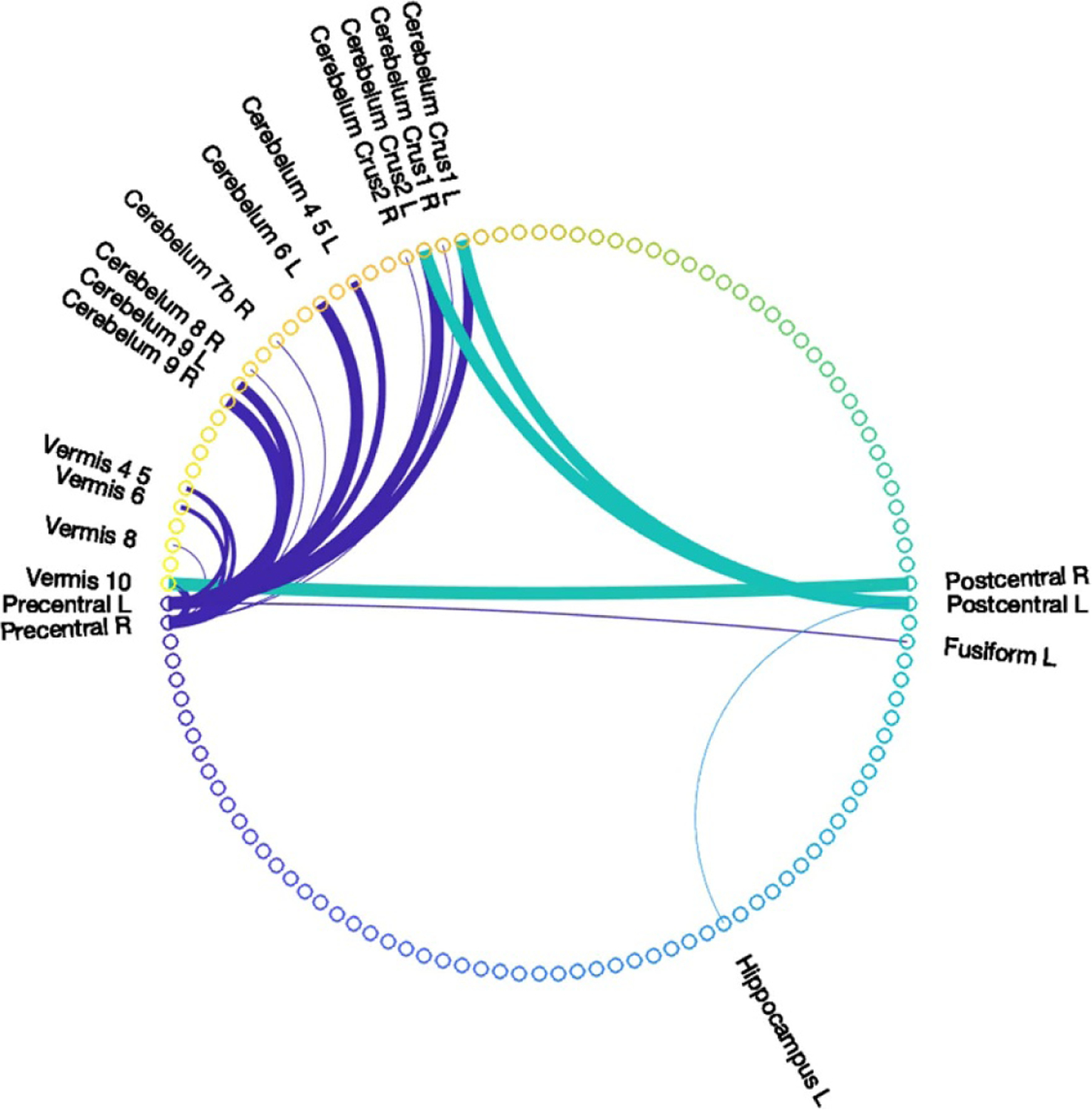
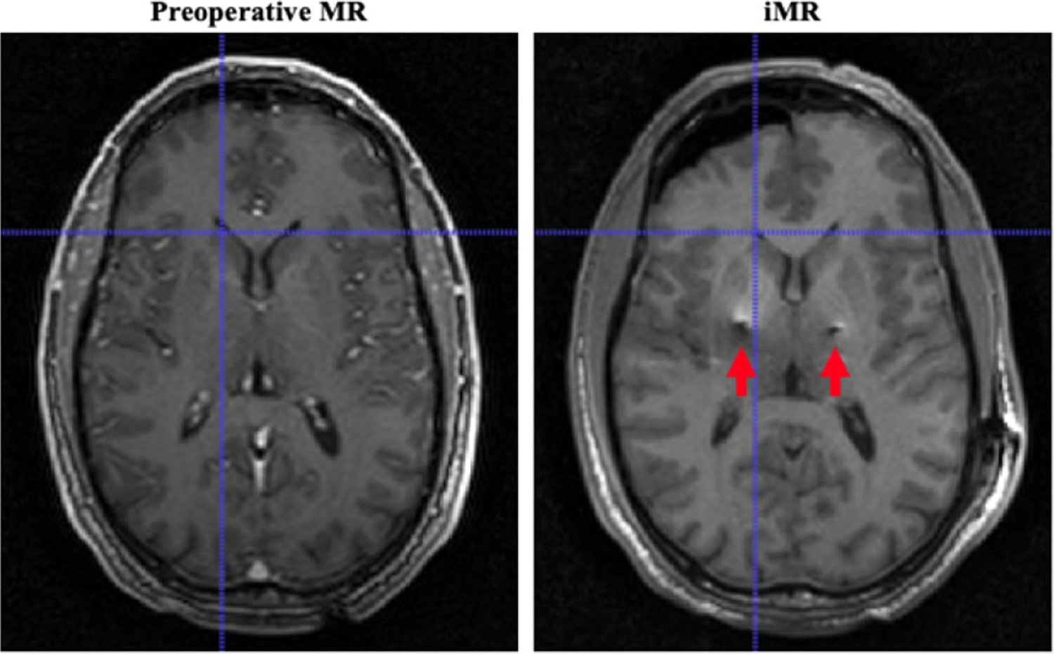
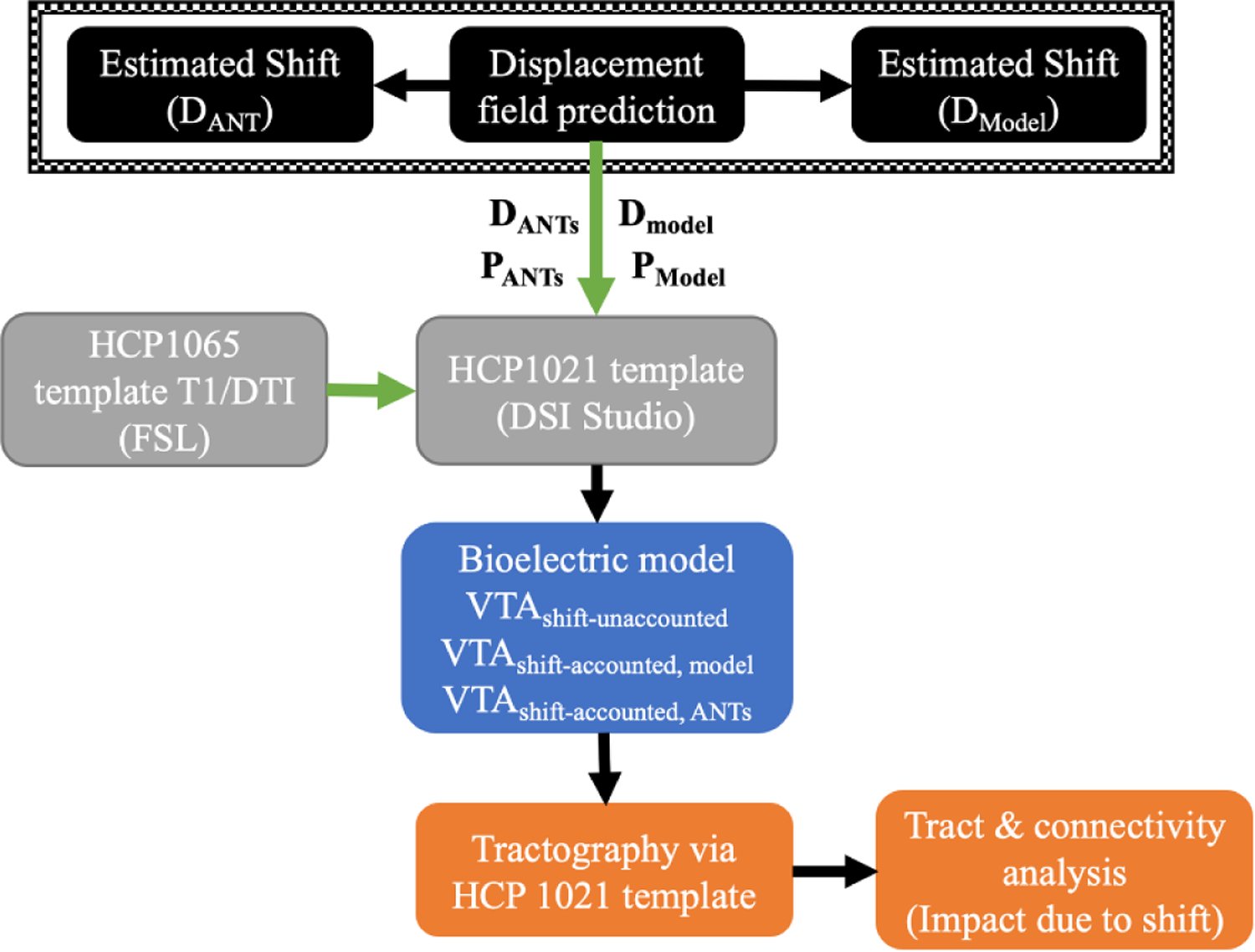
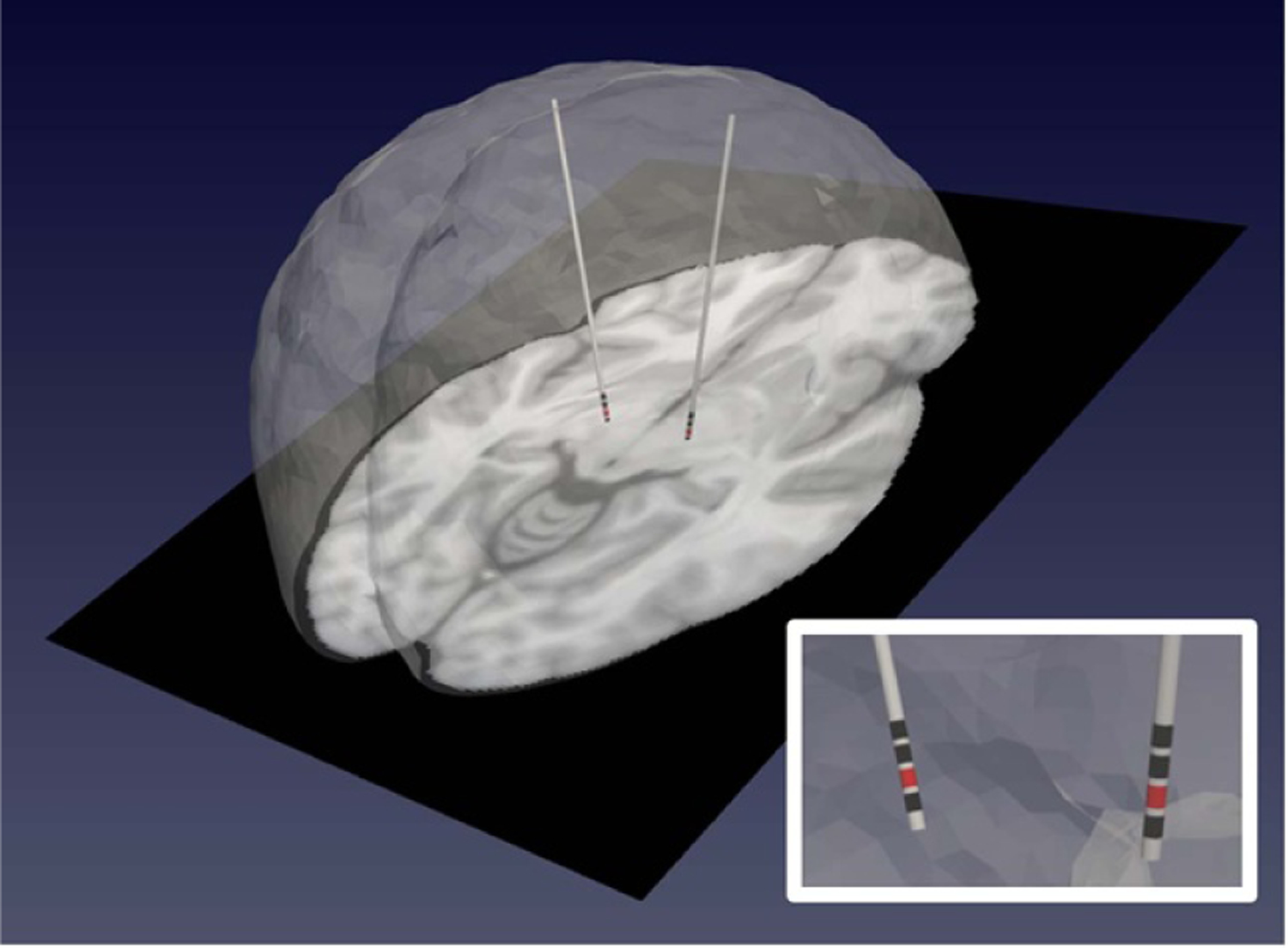
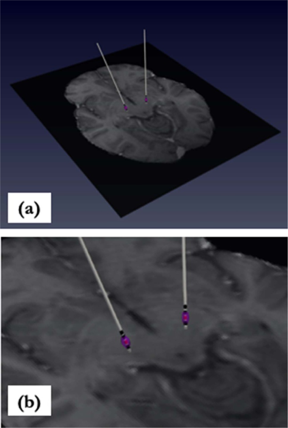
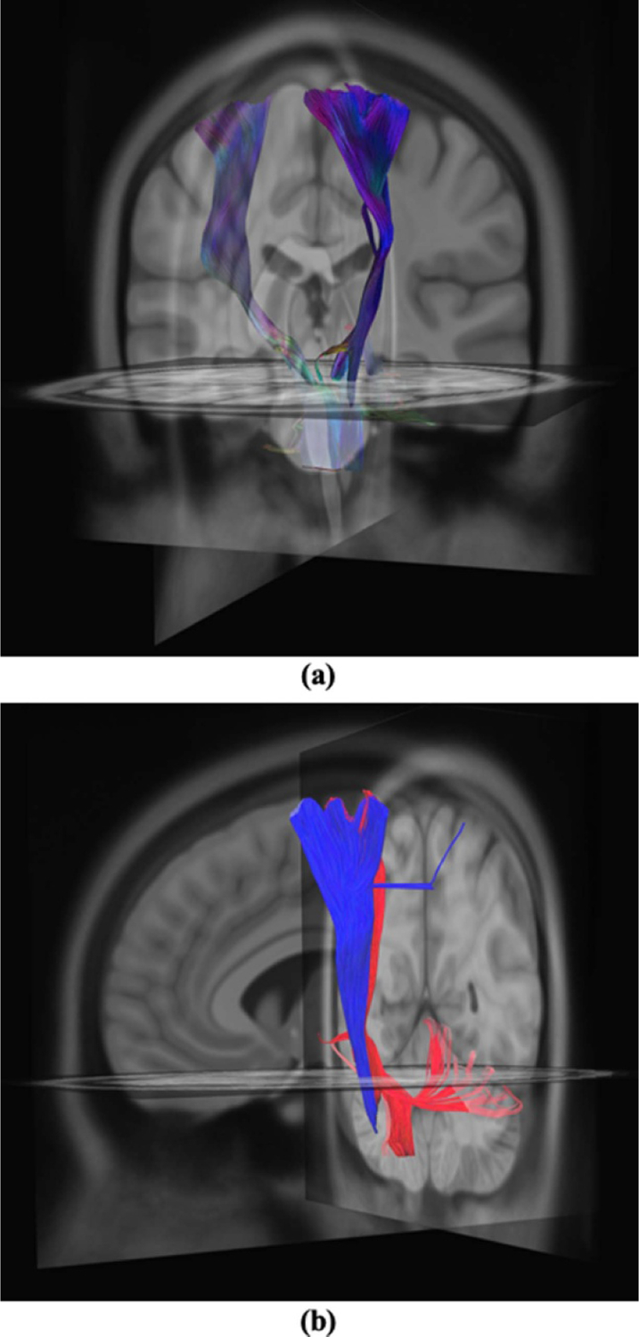
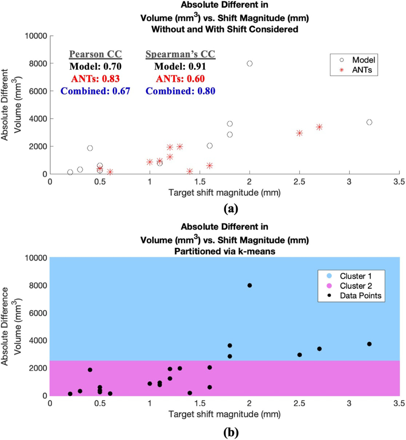
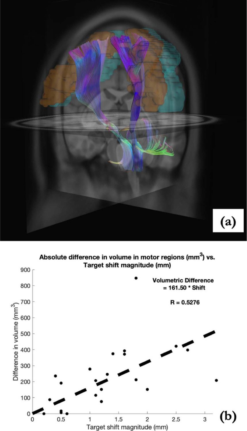
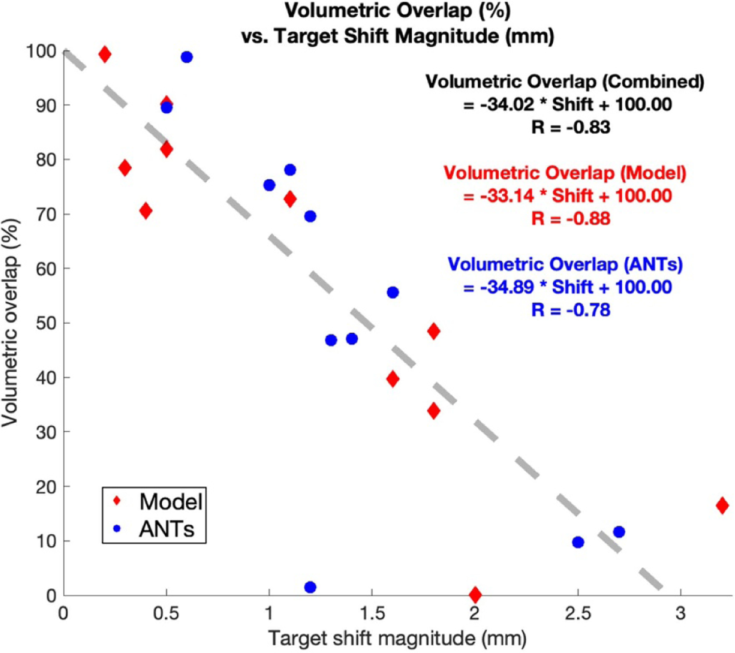
The effectiveness of deep brain stimulation (DBS) depends on electrode placement accuracy, which can be compromised by brain shift during surgery. While there have been efforts in assessing the impact of electrode misplacement due to brain shift using preop- and postop-imaging data, such analysis using preop- and intraop-imaging data via biophysical modeling has not been conducted. This work presents a preliminary study that applies a multi-physics analysis framework using finite element biomechanical and bioelectric models to examine the impact of realistic intraoperative shift on neural pathways determined by tractography.The study examined six patients who had undergone interventional magnetic resonance-guided DBS surgery. The modeling framework utilized a biomechanical approach to update preoperative MR to reflect shift-induced anatomical changes. Using this anatomically deformed image and its undeformed counterpart, bioelectric effects from shifting electrode leads could be simulated and neural activation differences were approximated. Specifically, for each configuration, volume of tissue activation was computed and subsequently used for tractography estimation. Total tract volume and overlapping volume with motor regions as well as connectivity profile were compared. In addition, volumetric overlap between different fiber bundles among configurations was computed and correlated to estimated shift.The study found deformation-induced differences in tract volume, motor region overlap, and connectivity behavior, suggesting the impact of shift. There is a strong correlation (= -0.83) between shift from intended target and intended neural pathway recruitment, where at threshold of ∼2.94 mm, intended recruitment completely degrades. The determined threshold is consistent with and provides quantitative support to prior observations and literature that deviations of 2-3 mm are detrimental.The findings support and advance prior studies and understanding to illustrate the need to account for shift in DBS and the potentiality of computational modeling for estimating influence of shift on neural activation.

摘要

深部脑刺激 (DBS) 的有效性取决于电极放置的准确性,而电极放置的准确性可能会因手术过程中的脑移位而受到影响。虽然已经有研究评估了由于脑移位导致的电极放置不当对术前和术后影像学数据的影响,但通过生物物理建模使用术前和术中影像学数据进行的这种分析尚未进行。这项工作提出了一项初步研究,该研究通过有限元生物力学和生物电模型的多物理分析框架,检查了通过示踪法确定的神经通路受现实术中移位影响的情况。该研究检查了六名接受介入性磁共振引导 DBS 手术的患者。建模框架利用生物力学方法更新术前磁共振成像,以反映移位引起的解剖结构变化。使用这种解剖变形图像及其未变形图像,可以模拟移位电极导线的生物电效应,并近似神经激活的差异。具体来说,对于每种配置,计算组织激活的体积,然后用于示踪法估计。比较了每种配置的激活体积、与运动区的重叠体积和连接性分布。此外,计算了不同配置之间不同纤维束之间的体积重叠,并将其与估计的移位相关联。该研究发现,在示踪体积、运动区重叠和连接行为方面存在变形引起的差异,这表明存在移位的影响。在从目标到目标神经通路募集的移位与目标之间存在很强的相关性(= -0.83),当达到约 2.94 毫米的阈值时,目标募集完全退化。确定的阈值与之前的观察结果和文献一致,并提供了定量支持,即 2-3 毫米的偏差是有害的。这些发现支持并推进了先前的研究和理解,以说明在 DBS 中考虑移位的必要性以及计算模型在估计移位对神经激活的影响方面的潜力。

https://cdn.ncbi.nlm.nih.gov/pmc/blobs/37ac/9476060/bbc99986c88c/nihms-1835634-f0011.jpg
https://cdn.ncbi.nlm.nih.gov/pmc/blobs/37ac/9476060/ed526497113f/nihms-1835634-f0002.jpg
https://cdn.ncbi.nlm.nih.gov/pmc/blobs/37ac/9476060/b295c3236790/nihms-1835634-f0003.jpg
https://cdn.ncbi.nlm.nih.gov/pmc/blobs/37ac/9476060/9a72d007ab88/nihms-1835634-f0004.jpg
https://cdn.ncbi.nlm.nih.gov/pmc/blobs/37ac/9476060/30094e50990b/nihms-1835634-f0005.jpg
https://cdn.ncbi.nlm.nih.gov/pmc/blobs/37ac/9476060/42d43d28b6ef/nihms-1835634-f0006.jpg
https://cdn.ncbi.nlm.nih.gov/pmc/blobs/37ac/9476060/f8071f937c52/nihms-1835634-f0007.jpg
https://cdn.ncbi.nlm.nih.gov/pmc/blobs/37ac/9476060/ec9721ce6f0a/nihms-1835634-f0008.jpg
https://cdn.ncbi.nlm.nih.gov/pmc/blobs/37ac/9476060/a4822ea28927/nihms-1835634-f0009.jpg
https://cdn.ncbi.nlm.nih.gov/pmc/blobs/37ac/9476060/538b2a7daef8/nihms-1835634-f0010.jpg
https://cdn.ncbi.nlm.nih.gov/pmc/blobs/37ac/9476060/bbc99986c88c/nihms-1835634-f0011.jpg