Trovato Piero, Simonetti Igino, Rinaldo Chiara, Grimaldi Dario, Verde Francesco, Lomoro Pascal, Codella Umberto, Rosa Ferdinando De, Corvino Antonio, Giovine Sabrina
Department of Radiology, SG Moscati Hospital, ASL Caserta, Aversa, Italy.
University of Naples Federico II, Department of Advanced Biomedical Sciences, Italy.
Pol J Radiol. 2021 Feb 1;86:e78-e86. doi: 10.5114/pjr.2021.103861. eCollection 2021.
To investigate the imaging features of emerging COVID-19 pneumonia on chest ultrasound, radiographs and computed tomography examinations performed at admission. In addition, we provide a review of the literature and compare our results with recent evidence regarding the imaging characteristics of this novel disease.
From March 17, 2020 to April 25, 2020, 23 patients with real-time polymerase chain reaction (RT-PCR) assay confirmed COVID-19 were identified. All 23 patients were evaluated and admitted at San Giuseppe Moscati Hospital in Aversa, Italy. Multi-modality imaging findings were evaluated and compared. Literature research was conducted through a methodical search on PubMed.
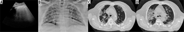
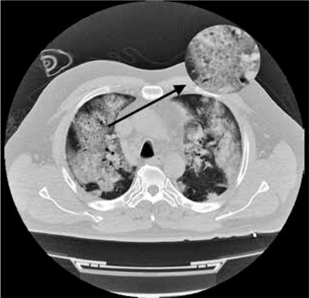
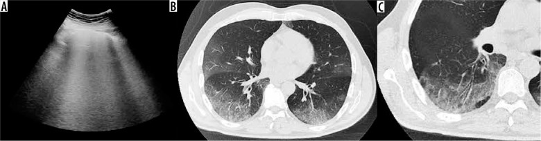
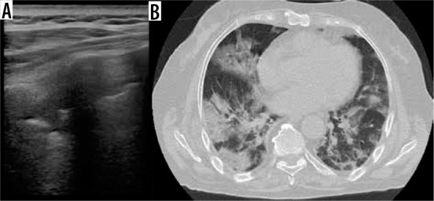
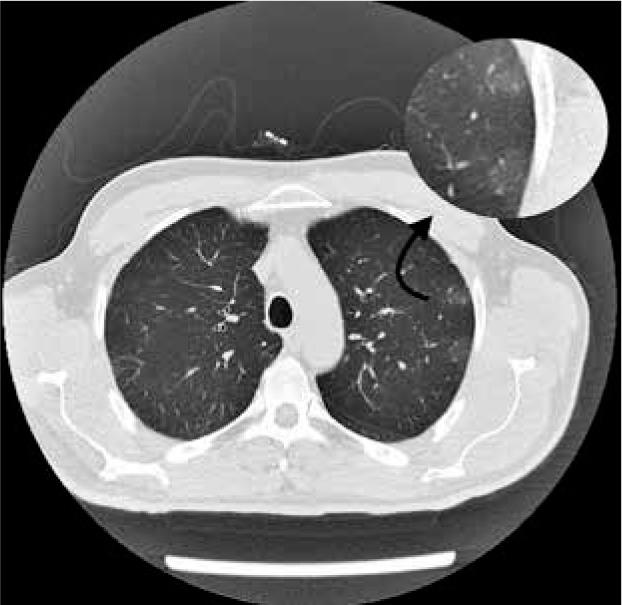
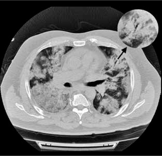
Twenty-three patients were included in the study. Chest transthoracic ultrasound (US), chest X-ray (CXR), and computed tomography (CT) were performed respectively in 11, 16 and 21 patients. Chest US findings were consistent with diffuse B lines (91%), subpleural consolidations (45%), and thickened pleural line (18%). CXR showed prevalent manifestations of consolidations (50%) and hazy increased opacities (37%). Typical CT features are bilateral and multilobar ground-glass opacities (GGO). Indeed GGO were present in 100% of our patients. Consolidations were visible in 76% of our study population. Notably both GGO and consolidations had a peripheral distribution in all our patients. Other CT imaging features included crazy-paving pattern, fibrous stripes, subpleural lines, architectural distortion, air bronchogram sign, vascular thickening and nodules. Our literature review identified thirty original studies supporting our imaging chest findings.
At admission, COVID-19 pneumonia can manifest in chest imaging as B-lines and consolidations on US, hazy opacities and consolidations on CXR, multiple GGO and consolidations on CT scan.
探讨新型冠状病毒肺炎(COVID-19)患者入院时胸部超声、X线及计算机断层扫描(CT)检查的影像学特征。此外,我们对相关文献进行综述,并将我们的结果与有关这种新型疾病影像学特征的最新证据进行比较。
2020年3月17日至2020年4月25日,共确诊23例实时荧光定量聚合酶链反应(RT-PCR)检测阳性的COVID-19患者。所有23例患者均在意大利阿韦尔萨的圣朱塞佩·莫斯卡蒂医院接受评估并入院治疗。对多模态影像学检查结果进行评估和比较。通过对PubMed进行系统检索开展文献研究。
本研究共纳入23例患者。分别对11例、16例和21例患者进行了胸部经胸超声(US)、胸部X线(CXR)和CT检查。胸部超声表现为弥漫性B线(91%)、胸膜下实变(45%)和胸膜线增厚(18%)。胸部X线显示实变(50%)和模糊的肺野透亮度增加(37%)为主要表现。典型的CT特征为双侧多叶磨玻璃影(GGO)。实际上,我们所有患者均出现GGO。76%的研究对象可见实变。值得注意的是,在我们所有患者中,GGO和实变均呈外周分布。其他CT影像学特征包括铺路石征、纤维条索、胸膜下线、结构扭曲、空气支气管征、血管增粗和结节。我们的文献综述确定了30项支持我们胸部影像学检查结果的原始研究。
入院时,COVID-19肺炎在胸部影像学上可表现为超声下的B线和实变、胸部X线下的模糊影和实变、CT扫描下的多发GGO和实变。