• 文献检索
  • 文档翻译
  • 深度研究
  • 学术资讯
  • Suppr Zotero 插件Zotero 插件
  • 邀请有礼
  • 套餐&价格
  • 历史记录
应用&插件
Suppr Zotero 插件Zotero 插件浏览器插件Mac 客户端Windows 客户端微信小程序
定价
高级版会员购买积分包购买API积分包
服务
文献检索文档翻译深度研究API 文档MCP 服务
关于我们
关于 Suppr公司介绍联系我们用户协议隐私条款
关注我们

Suppr 超能文献

核心技术专利:CN118964589B侵权必究
粤ICP备2023148730 号-1Suppr @ 2026

文献检索

告别复杂PubMed语法,用中文像聊天一样搜索,搜遍4000万医学文献。AI智能推荐,让科研检索更轻松。

立即免费搜索

文件翻译

保留排版,准确专业,支持PDF/Word/PPT等文件格式,支持 12+语言互译。

免费翻译文档

深度研究

AI帮你快速写综述,25分钟生成高质量综述,智能提取关键信息,辅助科研写作。

立即免费体验

新型冠状病毒肺炎的胸部薄层CT成像:轻型与重型患者的比较

Thin-Section Chest CT Imaging of COVID-19 Pneumonia: A Comparison Between Patients with Mild and Severe Disease.

作者信息

Yu Minhua, Xu Dan, Lan Lan, Tu Mengqi, Liao Rufang, Cai Shuhan, Cao Yiyuan, Xu Liying, Liao Meiyan, Zhang Xiaochun, Xiao Shu-Yuan, Li Yirong, Xu Haibo

机构信息

Departments of Radiology (M.Y., D.X., L.L., M.T., R.L., Y.C., L.X., M.L., X.Z., H.X.), Critical Care Medicine (S.C.), Pathology (S.Y.X.), and Laboratory Medicine (Y.L.), Zhongnan Hospital of Wuhan University, Wuchang District, Wuhan 430071, China; and Department of Pathology, University of Chicago Medicine, Chicago, Ill (S.Y.X.).

出版信息

Radiol Cardiothorac Imaging. 2020 Apr 23;2(2):e200126. doi: 10.1148/ryct.2020200126. eCollection 2020 Apr.

DOI:10.1148/ryct.2020200126
PMID:33778568
原文链接:https://pmc.ncbi.nlm.nih.gov/articles/PMC7233444/
Abstract

PURPOSE

To compare radiologic characteristics of coronavirus disease 2019 (COVID-19) pneumonia at thin-section CT on admission between patients with mild and severe disease.

MATERIALS AND METHODS

Seventy patients with COVID-19 pneumonia who were admitted to Zhongnan Hospital of Wuhan University between January 20, 2020 and January 27, 2020 were enrolled. On the basis of the World Health Organization guidelines, 50 patients were categorized with the mild form and 20 with the severe form based on clinical conditions. Imaging features, clinical, and laboratory data were reviewed and compared.

RESULTS

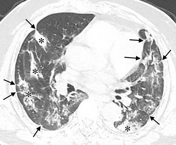
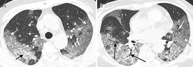
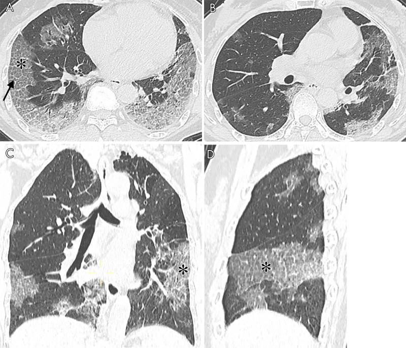
Patients with the severe form (median age, 65 years; interquartile range [IQR]: 54.75-75.00 years) were older than those with the mild form of disease (median age, 42.5 years; IQR: 32.75-58.50 years) ( < .001). Patients with the severe form of disease had more lung segments involved (median number of segments: 17.5 vs 7.5, ≤ .001) and also larger opacities (median number of segments with opacities measuring 3 cm to less than 50% of the lung segment: 5.5 vs 2.0, = .006; ≥ 50% of lung segment: 7.5 vs 0.0, < .001). They also had more interlobular septal thickening (75% vs 28%, < .001), higher prevalence of air bronchograms (70% vs 32%, = .004), and pleural effusions (40% vs 14%, = .017).

CONCLUSION

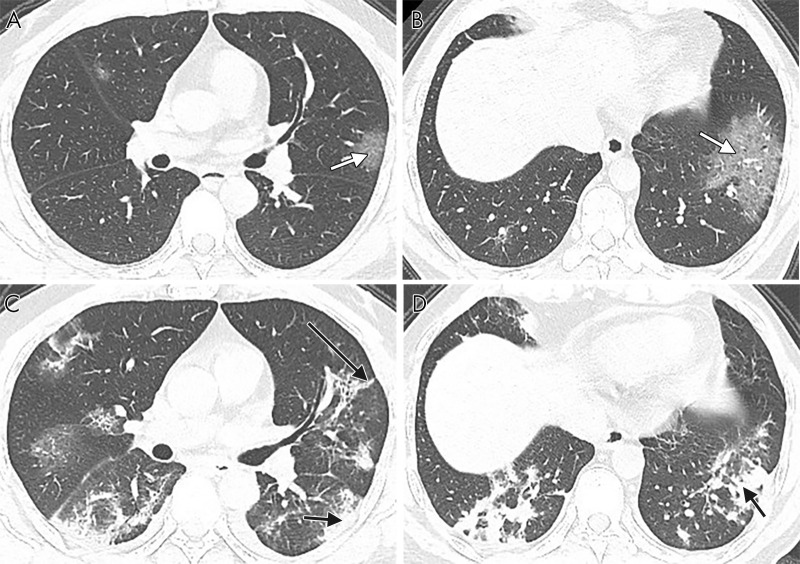
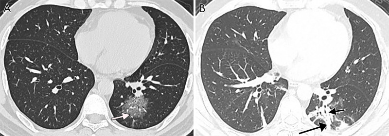
Ground-glass opacities with or without consolidation in a peripheral and basilar predominant distribution were the most common findings in COVID-19 pneumonia. Patients with the severe form of the disease had more extensive opacification of the lung parenchyma than did patients with mild disease. Interlobular septal thickening, air bronchograms, and pleural effusions were also more prevalent in severe COVID-19.© RSNA, 2020.

摘要

目的

比较2019冠状病毒病(COVID-19)肺炎患者入院时薄层CT的影像学特征,区分轻症和重症患者。

材料与方法

纳入2020年1月20日至1月27日期间在武汉大学中南医院收治的70例COVID-19肺炎患者。根据世界卫生组织指南,依据临床情况将50例患者归类为轻症,20例为重症。对影像学特征、临床和实验室数据进行回顾和比较。

结果

重症患者(中位年龄65岁;四分位间距[IQR]:54.75 - 75.00岁)比轻症患者(中位年龄42.5岁;IQR:32.75 - 58.50岁)年龄更大(P <.001)。重症患者累及的肺段更多(中位肺段数:17.5对7.5,P ≤.001),且实变更明显(直径3 cm至小于肺段50%的实变肺段中位数量:5.5对2.0,P =.006;≥肺段50%:7.5对0.0,P <.001)。他们还伴有更多的小叶间隔增厚(75%对28%,P <.001)、空气支气管征发生率更高(70%对32%,P =.004)以及胸腔积液(40%对14%,P =.017)。

结论

以磨玻璃影为主、有或无实变,以外周和基底部分布为主是COVID-19肺炎最常见的表现。重症患者肺实质的实变范围比轻症患者更广泛。小叶间隔增厚、空气支气管征和胸腔积液在重症COVID-19中也更常见。© RSNA,2020。

https://cdn.ncbi.nlm.nih.gov/pmc/blobs/d900/7977779/be4745c42671/ryct.2020200126.fig5.jpg
https://cdn.ncbi.nlm.nih.gov/pmc/blobs/d900/7977779/82976055ddff/ryct.2020200126.fig1.jpg
https://cdn.ncbi.nlm.nih.gov/pmc/blobs/d900/7977779/413a87cb5ff2/ryct.2020200126.fig2.jpg
https://cdn.ncbi.nlm.nih.gov/pmc/blobs/d900/7977779/f9de784de52d/ryct.2020200126.fig3.jpg
https://cdn.ncbi.nlm.nih.gov/pmc/blobs/d900/7977779/734a5ffb3866/ryct.2020200126.fig4.jpg
https://cdn.ncbi.nlm.nih.gov/pmc/blobs/d900/7977779/be4745c42671/ryct.2020200126.fig5.jpg

相似文献

1
Thin-Section Chest CT Imaging of COVID-19 Pneumonia: A Comparison Between Patients with Mild and Severe Disease.新型冠状病毒肺炎的胸部薄层CT成像:轻型与重型患者的比较
Radiol Cardiothorac Imaging. 2020 Apr 23;2(2):e200126. doi: 10.1148/ryct.2020200126. eCollection 2020 Apr.
2
COVID-19 pneumonia: CT findings of 122 patients and differentiation from influenza pneumonia.新型冠状病毒肺炎:122例患者的CT表现及与流感肺炎的鉴别诊断
Eur Radiol. 2020 Oct;30(10):5463-5469. doi: 10.1007/s00330-020-06928-0. Epub 2020 May 12.
3
Imaging features of the initial chest thin-section CT scans from 110 patients after admission with suspected or confirmed diagnosis of COVID-19.110 例疑似或确诊 COVID-19 患者入院后首次胸部薄层 CT 扫描的影像学特征。
BMC Med Imaging. 2020 Jun 15;20(1):64. doi: 10.1186/s12880-020-00464-5.
4
Imaging and clinical features of patients with 2019 novel coronavirus SARS-CoV-2.新型冠状病毒 2019 年 SARS-CoV-2 患者的影像学和临床特征。
Eur J Nucl Med Mol Imaging. 2020 May;47(5):1275-1280. doi: 10.1007/s00259-020-04735-9. Epub 2020 Feb 28.
5
Preliminary CT findings of coronavirus disease 2019 (COVID-19).新型冠状病毒肺炎(COVID-19)的初步 CT 影像学表现。
Clin Imaging. 2020 Sep;65:124-132. doi: 10.1016/j.clinimag.2020.04.042. Epub 2020 May 12.
6
COVID-19 pneumonia in the emergency department: correlation of initial chest CT findings with short-term outcome.急诊科的新型冠状病毒肺炎:初始胸部CT表现与短期预后的相关性
Emerg Radiol. 2020 Dec;27(6):691-699. doi: 10.1007/s10140-020-01863-5. Epub 2020 Oct 15.
7
A Comparison of Clinical and Chest CT Findings in Patients With Influenza A (H1N1) Virus Infection and Coronavirus Disease (COVID-19).甲型流感病毒(H1N1)感染与新型冠状病毒肺炎(COVID-19)患者的临床和胸部 CT 表现比较。
AJR Am J Roentgenol. 2020 Nov;215(5):1065-1071. doi: 10.2214/AJR.20.23214. Epub 2020 May 26.
8
Differential diagnosis between the coronavirus disease 2019 and Streptococcus pneumoniae pneumonia by thin-slice CT features.基于薄层 CT 特征的 2019 年冠状病毒病与肺炎链球菌肺炎的鉴别诊断。
Clin Imaging. 2021 Jan;69:318-323. doi: 10.1016/j.clinimag.2020.09.012. Epub 2020 Oct 6.
9
[Analysis of chest imaging features of novel coronavirus pneumonia, bacterial pneumonia and viral pneumonia].[新型冠状病毒肺炎、细菌性肺炎及病毒性肺炎的胸部影像特征分析]
Zhonghua Wei Zhong Bing Ji Jiu Yi Xue. 2023 Jan;35(1):28-31. doi: 10.3760/cma.j.cn121430-20210318-00394.
10
Chest CT-based differential diagnosis of 28 patients with suspected corona virus disease 2019 (COVID-19).基于胸部 CT 的 28 例疑似 2019 冠状病毒病(COVID-19)患者的鉴别诊断。
Br J Radiol. 2020 Aug;93(1112):20200243. doi: 10.1259/bjr.20200243. Epub 2020 Jun 2.

引用本文的文献

1
COVID-19 Computed tomography patterns in renal replacement therapy patients.COVID-19 患者在肾脏替代治疗中的计算机断层扫描模式。
J Bras Nefrol. 2024 Jul-Sep;46(3):e20230029. doi: 10.1590/2175-8239-JBN-2023-0029en.
2
Novel machine-learning analysis of SARS-CoV-2 infection in a subclinical nonhuman primate model using radiomics and blood biomarkers.利用放射组学和血液生物标志物对亚临床非人类灵长类动物模型中的 SARS-CoV-2 感染进行新型机器学习分析。
Sci Rep. 2023 Nov 10;13(1):19607. doi: 10.1038/s41598-023-46694-9.
3
Electrical Impedance Tomographic Mapping of Hypoventilated Lung Areas in Intubated Patients With COVID-19.

本文引用的文献

1
Severe COVID-19 Pneumonia: Assessing Inflammation Burden with Volume-rendered Chest CT.重症新型冠状病毒肺炎:利用容积再现胸部CT评估炎症负荷
Radiol Cardiothorac Imaging. 2020 Mar 6;2(2):e200044. doi: 10.1148/ryct.2020200044. eCollection 2020 Apr.
2
Clinical Characteristics of Coronavirus Disease 2019 in China.《中国 2019 年冠状病毒病临床特征》
N Engl J Med. 2020 Apr 30;382(18):1708-1720. doi: 10.1056/NEJMoa2002032. Epub 2020 Feb 28.
3
Initial CT findings and temporal changes in patients with the novel coronavirus pneumonia (2019-nCoV): a study of 63 patients in Wuhan, China.
新型冠状病毒肺炎(COVID-19)插管患者肺通气不足区域的电阻抗断层成像映射
Respir Care. 2023 Jun;68(6):773-776. doi: 10.4187/respcare.10261. Epub 2023 Apr 25.
4
Unilateral Pleural Effusion after Third Dose of BNT162b2 mRNA Vaccination: Case Report.BNT162b2 mRNA疫苗第三剂接种后单侧胸腔积液:病例报告
J Pers Med. 2023 Feb 23;13(3):391. doi: 10.3390/jpm13030391.
5
Abnormal Chest Computed Tomography Findings among Admitted Symptomatic COVID-19 Patients in a Tertiary Care Centre: A Descriptive Cross-sectional Study.收治的有症状 COVID-19 患者中异常胸部计算机断层扫描结果:一项描述性横断面研究。
JNMA J Nepal Med Assoc. 2022 Jul 1;60(251):608-611. doi: 10.31729/jnma.7529.
6
The role of Lung Ultrasound in the diagnosis of SARS-COV-2 disease in pregnant women.肺部超声在孕妇 SARS-COV-2 疾病诊断中的作用。
J Ultrasound. 2023 Jun;26(2):497-503. doi: 10.1007/s40477-022-00745-5. Epub 2022 Dec 27.
7
Significance of hematologic abnormalities in COVID-19 severity among infected patients in Lagos, Nigeria.尼日利亚拉各斯感染患者中血液学异常在新冠病毒病严重程度中的意义
Bull Natl Res Cent. 2022;46(1):275. doi: 10.1186/s42269-022-00959-x. Epub 2022 Dec 2.
8
Co-infection of SARS-CoV-2 with other respiratory pathogens in patients with liver disease.肝病患者中新型冠状病毒与其他呼吸道病原体的合并感染。
Access Microbiol. 2022 Oct 3;4(10):acmi000456. doi: 10.1099/acmi.0.000456. eCollection 2022.
9
CT Examinations for COVID-19: A Systematic Review of Protocols, Radiation Dose, and Numbers Needed to Diagnose and Predict.用于新型冠状病毒肺炎的CT检查:对方案、辐射剂量以及诊断和预测所需数量的系统评价
Taehan Yongsang Uihakhoe Chi. 2021 Nov;82(6):1505-1523. doi: 10.3348/jksr.2021.0096. Epub 2021 Nov 4.
10
The Caliber of Segmental and Subsegmental Vessels in COVID-19 Pneumonia Is Enlarged: A Distinctive Feature in Comparison with Other Forms of Inflammatory and Thromboembolic Diseases.新冠病毒肺炎节段性和亚节段性血管管径增宽:与其他形式的炎症性和血栓栓塞性疾病相比的一个显著特征。
J Pers Med. 2022 Sep 7;12(9):1465. doi: 10.3390/jpm12091465.
新型冠状病毒肺炎(2019-nCoV)患者的初始 CT 表现及时间演变:中国武汉 63 例患者研究。
Eur Radiol. 2020 Jun;30(6):3306-3309. doi: 10.1007/s00330-020-06731-x. Epub 2020 Feb 13.
4
Time Course of Lung Changes at Chest CT during Recovery from Coronavirus Disease 2019 (COVID-19).新冠肺炎(COVID-19)康复过程中胸部 CT 肺部变化的时间进程。
Radiology. 2020 Jun;295(3):715-721. doi: 10.1148/radiol.2020200370. Epub 2020 Feb 13.
5
What to do next to control the 2019-nCoV epidemic?接下来要采取什么措施来控制2019新型冠状病毒疫情?
Lancet. 2020 Feb 8;395(10222):391-393. doi: 10.1016/S0140-6736(20)30300-7.
6
Clinical Characteristics of 138 Hospitalized Patients With 2019 Novel Coronavirus-Infected Pneumonia in Wuhan, China.《武汉 2019 年新型冠状病毒感染的肺炎 138 例住院患者临床特征分析》
JAMA. 2020 Mar 17;323(11):1061-1069. doi: 10.1001/jama.2020.1585.
7
Evolving status of the 2019 novel coronavirus infection: Proposal of conventional serologic assays for disease diagnosis and infection monitoring.2019新型冠状病毒感染的演变情况:疾病诊断和感染监测常规血清学检测方法的建议
J Med Virol. 2020 May;92(5):464-467. doi: 10.1002/jmv.25702. Epub 2020 Feb 17.
8
Emerging 2019 Novel Coronavirus (2019-nCoV) Pneumonia.新型冠状病毒(2019-nCoV)肺炎的诊治进展。
Radiology. 2020 Apr;295(1):210-217. doi: 10.1148/radiol.2020200274. Epub 2020 Feb 6.
9
CT Imaging Features of 2019 Novel Coronavirus (2019-nCoV).2019 新型冠状病毒(2019-nCoV)的 CT 影像学特征。
Radiology. 2020 Apr;295(1):202-207. doi: 10.1148/radiol.2020200230. Epub 2020 Feb 4.
10
Epidemiological and clinical characteristics of 99 cases of 2019 novel coronavirus pneumonia in Wuhan, China: a descriptive study.中国武汉 99 例 2019 年新型冠状病毒肺炎患者的流行病学和临床特征:描述性研究。
Lancet. 2020 Feb 15;395(10223):507-513. doi: 10.1016/S0140-6736(20)30211-7. Epub 2020 Jan 30.