• 文献检索
  • 文档翻译
  • 深度研究
  • 学术资讯
  • Suppr Zotero 插件Zotero 插件
  • 邀请有礼
  • 套餐&价格
  • 历史记录
应用&插件
Suppr Zotero 插件Zotero 插件浏览器插件Mac 客户端Windows 客户端微信小程序
定价
高级版会员购买积分包购买API积分包
服务
文献检索文档翻译深度研究API 文档MCP 服务
关于我们
关于 Suppr公司介绍联系我们用户协议隐私条款
关注我们

Suppr 超能文献

核心技术专利:CN118964589B侵权必究
粤ICP备2023148730 号-1Suppr @ 2026

文献检索

告别复杂PubMed语法,用中文像聊天一样搜索,搜遍4000万医学文献。AI智能推荐,让科研检索更轻松。

立即免费搜索

文件翻译

保留排版,准确专业,支持PDF/Word/PPT等文件格式,支持 12+语言互译。

免费翻译文档

深度研究

AI帮你快速写综述,25分钟生成高质量综述,智能提取关键信息,辅助科研写作。

立即免费体验

圆锥角膜与正常眼角膜表面面积的定量比较。

Quantitative comparison of corneal surface areas in keratoconus and normal eyes.

机构信息

Rothschild Foundation Hospital, Paris, France.

CHR Citadelle, Liège, Belgique.

出版信息

Sci Rep. 2021 Mar 25;11(1):6840. doi: 10.1038/s41598-021-86185-3.

DOI:10.1038/s41598-021-86185-3
PMID:33767220
原文链接:https://pmc.ncbi.nlm.nih.gov/articles/PMC7994394/
Abstract

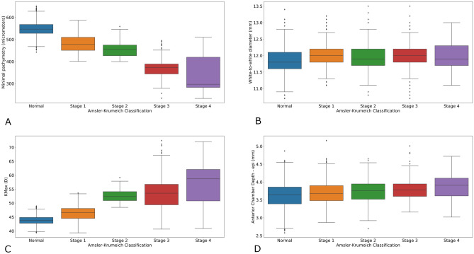
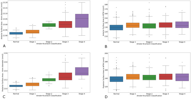
Keratoconus is a highly prevalent corneal disorder characterized by progressive corneal thinning, steepening and irregular astigmatism. To date, pathophysiology of keratoconus development and progression remains debated. In this study, we retrospectively analysed topographic elevation maps from 3227 eyes of 3227 patients (969 keratoconus and 2258 normal eyes) to calculate anterior and posterior corneal surface area. We compared results from normal eyes and keratoconus eyes using the Mann-Whitney U test. The Kruskal-Wallis test was used to compare keratoconus stages according to the Amsler-Krumeich classification. Keratoconus eyes were shown to have statistically significantly larger corneal surface areas, measured at the central 4.0 mm and 8.0 mm, and total corneal diameter. However, no significant increase in corneal surface area was seen with increasing severity of keratoconus. We suggest that these results indicate redistribution, rather than increase, of the corneal surface area with keratoconus severity.

摘要

圆锥角膜是一种常见的角膜疾病,其特征为进行性角膜变薄、变陡和不规则散光。迄今为止,圆锥角膜的发病机制和进展仍存在争议。在这项研究中,我们回顾性地分析了 3227 名患者(969 名圆锥角膜和 2258 名正常眼)的 3227 只眼的地形图隆起图,以计算前、后角膜表面积。我们使用曼-惠特尼 U 检验比较了正常眼和圆锥角膜眼的结果。Kruskal-Wallis 检验用于根据 Amsler-Krumeich 分类比较圆锥角膜的分期。结果表明,在中央 4.0mm 和 8.0mm 以及总角膜直径处,圆锥角膜眼的角膜表面积明显更大,且具有统计学意义。然而,随着圆锥角膜严重程度的增加,角膜表面积并没有明显增加。我们认为,这些结果表明,随着圆锥角膜严重程度的增加,角膜表面积的分布发生了变化,而不是增加。

https://cdn.ncbi.nlm.nih.gov/pmc/blobs/1f5c/7994394/6b5e0a4f8d79/41598_2021_86185_Fig4_HTML.jpg
https://cdn.ncbi.nlm.nih.gov/pmc/blobs/1f5c/7994394/0884a9d73eb9/41598_2021_86185_Fig1_HTML.jpg
https://cdn.ncbi.nlm.nih.gov/pmc/blobs/1f5c/7994394/edb1a7ef2be8/41598_2021_86185_Fig2_HTML.jpg
https://cdn.ncbi.nlm.nih.gov/pmc/blobs/1f5c/7994394/11924bdc0a50/41598_2021_86185_Fig3_HTML.jpg
https://cdn.ncbi.nlm.nih.gov/pmc/blobs/1f5c/7994394/6b5e0a4f8d79/41598_2021_86185_Fig4_HTML.jpg

相似文献

1
Quantitative comparison of corneal surface areas in keratoconus and normal eyes.圆锥角膜与正常眼角膜表面面积的定量比较。
Sci Rep. 2021 Mar 25;11(1):6840. doi: 10.1038/s41598-021-86185-3.
2
Involvement of anterior and posterior corneal surface area imbalance in the pathological change of keratoconus.角膜前表面和后表面区域失衡在圆锥角膜病变中的作用。
Sci Rep. 2018 Oct 9;8(1):14993. doi: 10.1038/s41598-018-33490-z.
3
Correlation of corneal elevation with severity of keratoconus by means of anterior and posterior topographic analysis.通过前、后表面地形图分析角膜隆起与圆锥角膜严重程度的相关性。
Cornea. 2012 Mar;31(3):253-8. doi: 10.1097/ICO.0B013E31823D1EE0.
4
Corneal differences between healthy and subclinical patients assessed using two diferente corneal tomographers.使用两种不同的角膜地形图仪评估健康与亚临床患者之间的角膜差异。
Arq Bras Oftalmol. 2020 Mar-Apr;83(2):92-97. doi: 10.5935/0004-2749.20200015.
5
The variability of corneal and anterior segment parameters in keratoconus.圆锥角膜中角膜和眼前节参数的变异性。
Cont Lens Anterior Eye. 2016 Dec;39(6):466-470. doi: 10.1016/j.clae.2016.05.003. Epub 2016 Jun 1.
6
Characteristics of Corneal Astigmatism of Anterior and Posterior Surface in a Normal Control Group and Patients With Keratoconus.正常对照组和圆锥角膜患者前后表面角膜散光的特征
Cornea. 2017 Apr;36(4):457-462. doi: 10.1097/ICO.0000000000001143.
7
Quantitative interocular comparison of total corneal surface area and corneal diameter in patients with highly asymmetric keratoconus.高度非对称圆锥角膜患者的全角膜表面面积和角膜直径的定量眼间比较。
Sci Rep. 2022 Mar 11;12(1):4276. doi: 10.1038/s41598-022-08021-6.
8
[Analysis on the role of Sirius combined topography and tomography system in screening for suspect keratoconus].[Sirius综合地形和断层扫描系统在圆锥角膜疑似病例筛查中的作用分析]
Zhonghua Yan Ke Za Zhi. 2018 Jan 11;54(1):33-38. doi: 10.3760/cma.j.issn.0412-4081.2018.01.007.
9
Corneal higher order aberrations: a method to grade keratoconus.角膜高阶像差:一种圆锥角膜分级方法。
J Refract Surg. 2006 Jun;22(6):539-45. doi: 10.3928/1081-597X-20060601-05.
10
Keratoconus staging with ultrasound biomicroscopy.超声生物显微镜下圆锥角膜分期
Ophthalmologica. 1998;212 Suppl 1:10-2. doi: 10.1159/000055412.

引用本文的文献

1
Corneal surface changes after stromal lenticule addition keratoplasty combined with cross-linking for severe keratoconus.基质透镜植入角膜移植联合交联治疗重度圆锥角膜后的角膜表面变化
Int J Ophthalmol. 2025 Jun 18;18(6):1003-1010. doi: 10.18240/ijo.2025.06.05. eCollection 2025.
2
Measurements of white-to-white corneal diameter and anterior chamber parameters using the Pentacam AXL wave and their correlations in the adult Saudi population.使用Pentacam AXL波测量沙特成年人群的角膜白对白直径和前房参数及其相关性
PeerJ. 2025 Apr 1;13:e19227. doi: 10.7717/peerj.19227. eCollection 2025.
3
Topographic factors associated with anterior chamber angle narrowing in patients with keratoconus.

本文引用的文献

1
Corneal Topography Raw Data Classification Using a Convolutional Neural Network.基于卷积神经网络的角膜地形原始数据分类。
Am J Ophthalmol. 2020 Nov;219:33-39. doi: 10.1016/j.ajo.2020.06.005. Epub 2020 Jun 10.
2
Anterior and posterior ratio of corneal surface areas: A novel index for detecting early stage keratoconus.角膜表面面积的前后比:一种用于早期检测圆锥角膜的新指标。
PLoS One. 2020 Apr 2;15(4):e0231074. doi: 10.1371/journal.pone.0231074. eCollection 2020.
3
Involvement of anterior and posterior corneal surface area imbalance in the pathological change of keratoconus.
圆锥角膜患者前房角狭窄相关的地形学因素。
Int J Ophthalmol. 2025 Jan 18;18(1):67-73. doi: 10.18240/ijo.2025.01.07. eCollection 2025.
4
Keratoconus Progression Determined at the First Visit: A Deep Learning Approach With Fusion of Imaging and Numerical Clinical Data.初次就诊时确定的圆锥角膜进展:融合成像和数值临床数据的深度学习方法。
Transl Vis Sci Technol. 2024 May 1;13(5):7. doi: 10.1167/tvst.13.5.7.
5
Use of Nanoindentation in Determination of Regional Biomechanical Properties of Rabbit Cornea After UVA Cross-Linking.纳米压痕法在评估 UVA 交联后兔眼角膜区域性生物力学性能中的应用。
Invest Ophthalmol Vis Sci. 2023 Oct 3;64(13):26. doi: 10.1167/iovs.64.13.26.
6
Quantitative interocular comparison of total corneal surface area and corneal diameter in patients with highly asymmetric keratoconus.高度非对称圆锥角膜患者的全角膜表面面积和角膜直径的定量眼间比较。
Sci Rep. 2022 Mar 11;12(1):4276. doi: 10.1038/s41598-022-08021-6.
7
Interocular Symmetry Analysis of Corneal Elevation Using the Fellow Eye as the Reference Surface and Machine Learning.以对侧眼为参考面并运用机器学习的角膜高度眼间对称性分析
Healthcare (Basel). 2021 Dec 16;9(12):1738. doi: 10.3390/healthcare9121738.
角膜前表面和后表面区域失衡在圆锥角膜病变中的作用。
Sci Rep. 2018 Oct 9;8(1):14993. doi: 10.1038/s41598-018-33490-z.
4
Evaluation of keratoconus progression.评估圆锥角膜的进展。
Br J Ophthalmol. 2019 Apr;103(4):551-557. doi: 10.1136/bjophthalmol-2017-311651. Epub 2018 Jun 1.
5
A new approach to keratoconus detection based on corneal morphogeometric analysis.一种基于角膜形态几何分析的圆锥角膜检测新方法。
PLoS One. 2017 Sep 8;12(9):e0184569. doi: 10.1371/journal.pone.0184569. eCollection 2017.
6
Repeatability of Keratometry Measurements Obtained With Three Topographers in Keratoconic and Normal Corneas.三种角膜地形图仪在圆锥角膜和正常角膜上获得的角膜曲率测量的可重复性
J Refract Surg. 2016 Mar;32(3):187-92. doi: 10.3928/1081597X-20160113-01.
7
Global consensus on keratoconus and ectatic diseases.圆锥角膜和扩张性疾病的全球共识。
Cornea. 2015 Apr;34(4):359-69. doi: 10.1097/ICO.0000000000000408.
8
Geometrical custom modeling of human cornea in vivo and its use for the diagnosis of corneal ectasia.人眼角膜活体几何定制建模及其在圆锥角膜诊断中的应用。
PLoS One. 2014 Oct 17;9(10):e110249. doi: 10.1371/journal.pone.0110249. eCollection 2014.
9
Biomechanics of corneal ectasia and biomechanical treatments.角膜扩张症的生物力学与生物力学治疗
J Cataract Refract Surg. 2014 Jun;40(6):991-8. doi: 10.1016/j.jcrs.2014.04.013. Epub 2014 Apr 26.
10
What causes keratoconus?是什么导致了圆锥角膜?
Cornea. 2012 Jun;31(6):716-9. doi: 10.1097/ICO.0b013e31823f8c72.