Department of Radiological Sciences, David Geffen School of Medicine at University of California Los Angeles, 757 Westwood Plaza, Suite 1621, Los Angeles, CA, 90095-7532, USA.
Department of Diagnostic, Molecular and Interventional Radiology, Icahn School of Medicine at Mount Sinai, New York, USA.
Sci Rep. 2021 Mar 25;11(1):6876. doi: 10.1038/s41598-021-86022-7.
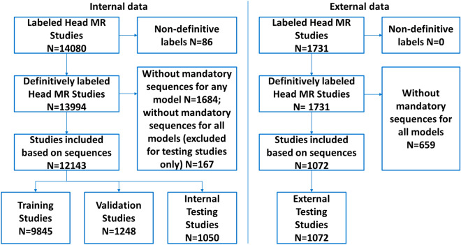
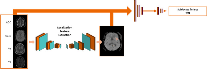
With the rapid growth and increasing use of brain MRI, there is an interest in automated image classification to aid human interpretation and improve workflow. We aimed to train a deep convolutional neural network and assess its performance in identifying abnormal brain MRIs and critical intracranial findings including acute infarction, acute hemorrhage and mass effect. A total of 13,215 clinical brain MRI studies were categorized to training (74%), validation (9%), internal testing (8%) and external testing (8%) datasets. Up to eight contrasts were included from each brain MRI and each image volume was reformatted to common resolution to accommodate for differences between scanners. Following reviewing the radiology reports, three neuroradiologists assigned each study to abnormal vs normal, and identified three critical findings including acute infarction, acute hemorrhage, and mass effect. A deep convolutional neural network was constructed by a combination of localization feature extraction (LFE) modules and global classifiers to identify the presence of 4 variables in brain MRIs including abnormal, acute infarction, acute hemorrhage and mass effect. Training, validation and testing sets were randomly defined on a patient basis. Training was performed on 9845 studies using balanced sampling to address class imbalance. Receiver operating characteristic (ROC) analysis was performed. The ROC analysis of our models for 1050 studies within our internal test data showed AUC/sensitivity/specificity of 0.91/83%/86% for normal versus abnormal brain MRI, 0.95/92%/88% for acute infarction, 0.90/89%/81% for acute hemorrhage, and 0.93/93%/85% for mass effect. For 1072 studies within our external test data, it showed AUC/sensitivity/specificity of 0.88/80%/80% for normal versus abnormal brain MRI, 0.97/90%/97% for acute infarction, 0.83/72%/88% for acute hemorrhage, and 0.87/79%/81% for mass effect. Our proposed deep convolutional network can accurately identify abnormal and critical intracranial findings on individual brain MRIs, while addressing the fact that some MR contrasts might not be available in individual studies.
随着脑 MRI 的快速增长和日益普及,人们对自动图像分类产生了兴趣,以辅助人工解读并提高工作流程效率。我们旨在训练一个深度卷积神经网络,并评估其在识别异常脑 MRI 和关键颅内发现(包括急性梗死、急性出血和占位效应)方面的性能。总共将 13215 项临床脑部 MRI 研究分为训练集(74%)、验证集(9%)、内部测试集(8%)和外部测试集(8%)。每个脑部 MRI 包含多达 8 个对比度,每个图像体积都被重新格式化到常见的分辨率,以适应扫描仪之间的差异。在查看放射学报告后,三位神经放射学家将每个研究分配为异常与正常,并确定了三个关键发现,包括急性梗死、急性出血和占位效应。通过组合定位特征提取(LFE)模块和全局分类器构建了一个深度卷积神经网络,以识别脑部 MRI 中 4 个变量(异常、急性梗死、急性出血和占位效应)的存在情况。训练、验证和测试集是基于患者随机定义的。在 9845 项研究中使用平衡采样进行训练,以解决类别不平衡问题。进行了接收者操作特征(ROC)分析。我们的模型在内部测试数据的 1050 项研究中的 ROC 分析显示,正常与异常脑部 MRI 的 AUC/敏感性/特异性为 0.91/83%/86%,急性梗死为 0.95/92%/88%,急性出血为 0.90/89%/81%,占位效应为 0.93/93%/85%。对于外部测试数据中的 1072 项研究,其结果显示正常与异常脑部 MRI 的 AUC/敏感性/特异性为 0.88/80%/80%,急性梗死为 0.97/90%/97%,急性出血为 0.83/72%/88%,占位效应为 0.87/79%/81%。我们提出的深度卷积网络可以准确识别个体脑部 MRI 上的异常和关键颅内发现,同时解决了某些 MRI 对比度在个别研究中可能不可用的问题。