• 文献检索
  • 文档翻译
  • 深度研究
  • 学术资讯
  • Suppr Zotero 插件Zotero 插件
  • 邀请有礼
  • 套餐&价格
  • 历史记录
应用&插件
Suppr Zotero 插件Zotero 插件浏览器插件Mac 客户端Windows 客户端微信小程序
定价
高级版会员购买积分包购买API积分包
服务
文献检索文档翻译深度研究API 文档MCP 服务
关于我们
关于 Suppr公司介绍联系我们用户协议隐私条款
关注我们

Suppr 超能文献

核心技术专利:CN118964589B侵权必究
粤ICP备2023148730 号-1Suppr @ 2026

文献检索

告别复杂PubMed语法,用中文像聊天一样搜索,搜遍4000万医学文献。AI智能推荐,让科研检索更轻松。

立即免费搜索

文件翻译

保留排版,准确专业,支持PDF/Word/PPT等文件格式,支持 12+语言互译。

免费翻译文档

深度研究

AI帮你快速写综述,25分钟生成高质量综述,智能提取关键信息,辅助科研写作。

立即免费体验

不同种族皮肤色素性疾病的皮肤镜特征

Dermatoscopic Features of Pigmentary Diseases in Ethnic Skin.

作者信息

Vinay Keshavamurthy, Ankad Balachandra S

机构信息

Department of Dermatology, Venereology and Leprology, Postgraduate Institute of Medical Education and Research, Chandigarh, Karnataka, India.

Department of Dermatology, S Nijalingappa Medical College, Bagalkot, Karnataka, India.

出版信息

Indian Dermatol Online J. 2021 Jan 16;12(1):24-33. doi: 10.4103/idoj.IDOJ_561_20. eCollection 2021 Jan-Feb.

DOI:10.4103/idoj.IDOJ_561_20
PMID:33768020
原文链接:https://pmc.ncbi.nlm.nih.gov/articles/PMC7982038/
Abstract

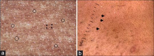
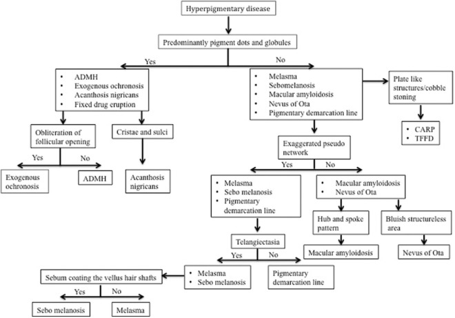
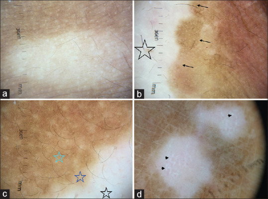
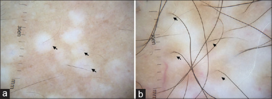
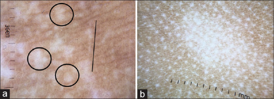
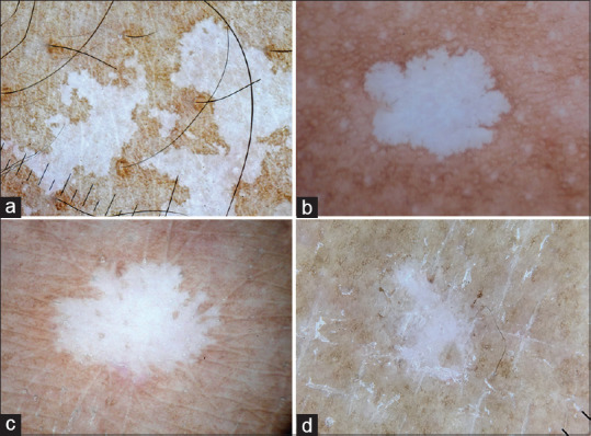
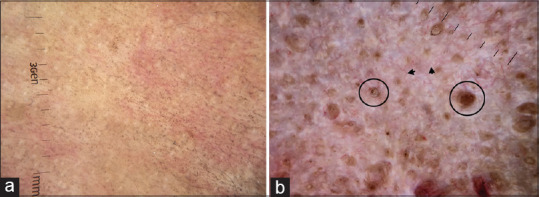
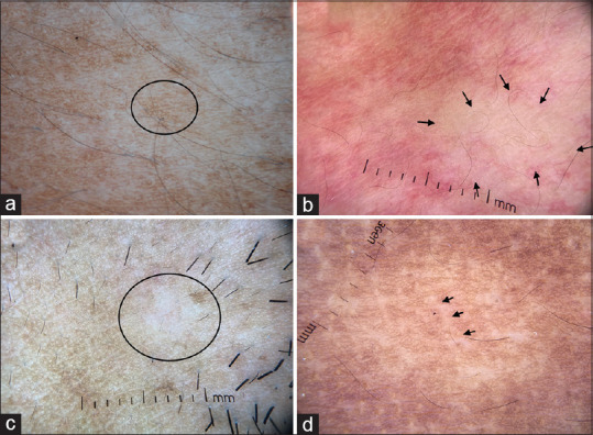
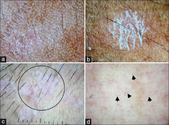
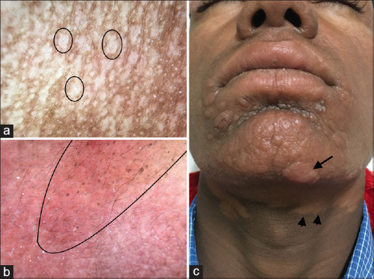
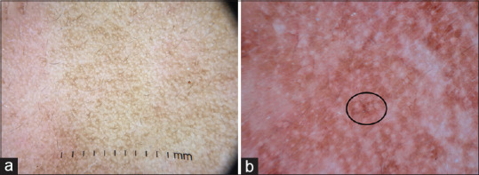
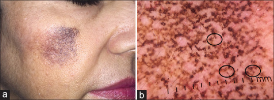
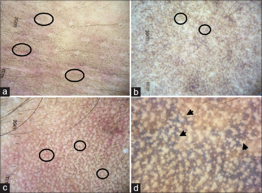
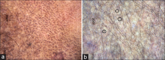
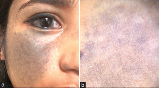
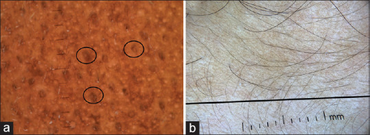
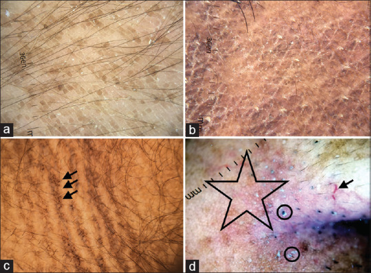
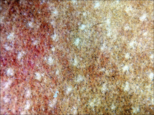
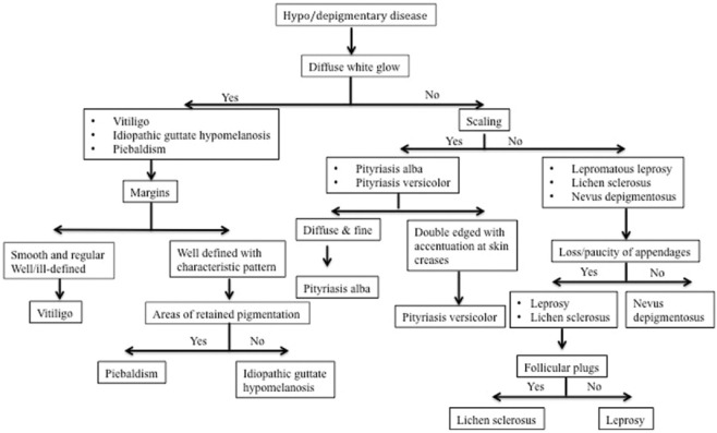
Dermatoscopy is a non-invasive, handy tool, which is increasingly being used in diagnosis and prognostication of pigmentary dermatoses. Dermatoscopic changes in pigmentary pattern, scaling, and vasculature help us to differentiate among the myriad of hypo and hyper pigmentary diseases. This review gives a brief overview of the dermatoscopic features of pigmentary diseases, which are commonly encountered in clinical practice. We also provide a diagnostic approach based on salient dermatoscopic features.

摘要

皮肤镜检查是一种非侵入性的便捷工具,越来越多地用于色素性皮肤病的诊断和预后评估。色素沉着模式、鳞屑和血管系统的皮肤镜改变有助于我们区分众多色素减退和色素沉着性疾病。本文综述了临床实践中常见色素性疾病的皮肤镜特征,并基于显著的皮肤镜特征提供了一种诊断方法。

https://cdn.ncbi.nlm.nih.gov/pmc/blobs/3ccc/7982038/29524b5f2654/IDOJ-12-24-g020.jpg
https://cdn.ncbi.nlm.nih.gov/pmc/blobs/3ccc/7982038/683fbd6b85fd/IDOJ-12-24-g001.jpg
https://cdn.ncbi.nlm.nih.gov/pmc/blobs/3ccc/7982038/aa1266c5a4a9/IDOJ-12-24-g002.jpg
https://cdn.ncbi.nlm.nih.gov/pmc/blobs/3ccc/7982038/7ef4f620d4f6/IDOJ-12-24-g003.jpg
https://cdn.ncbi.nlm.nih.gov/pmc/blobs/3ccc/7982038/c3f5ee1429cf/IDOJ-12-24-g004.jpg
https://cdn.ncbi.nlm.nih.gov/pmc/blobs/3ccc/7982038/2096dd1d0b0f/IDOJ-12-24-g005.jpg
https://cdn.ncbi.nlm.nih.gov/pmc/blobs/3ccc/7982038/5ab4d0448879/IDOJ-12-24-g006.jpg
https://cdn.ncbi.nlm.nih.gov/pmc/blobs/3ccc/7982038/1a2c700e2930/IDOJ-12-24-g007.jpg
https://cdn.ncbi.nlm.nih.gov/pmc/blobs/3ccc/7982038/75f6fa6cc8c9/IDOJ-12-24-g008.jpg
https://cdn.ncbi.nlm.nih.gov/pmc/blobs/3ccc/7982038/6e6ca8bfa207/IDOJ-12-24-g009.jpg
https://cdn.ncbi.nlm.nih.gov/pmc/blobs/3ccc/7982038/512a9117150a/IDOJ-12-24-g010.jpg
https://cdn.ncbi.nlm.nih.gov/pmc/blobs/3ccc/7982038/39733961ae8e/IDOJ-12-24-g011.jpg
https://cdn.ncbi.nlm.nih.gov/pmc/blobs/3ccc/7982038/afdfe6c6192e/IDOJ-12-24-g012.jpg
https://cdn.ncbi.nlm.nih.gov/pmc/blobs/3ccc/7982038/d8b5eae935ff/IDOJ-12-24-g013.jpg
https://cdn.ncbi.nlm.nih.gov/pmc/blobs/3ccc/7982038/7b95e20677e7/IDOJ-12-24-g014.jpg
https://cdn.ncbi.nlm.nih.gov/pmc/blobs/3ccc/7982038/02cac3eae10f/IDOJ-12-24-g015.jpg
https://cdn.ncbi.nlm.nih.gov/pmc/blobs/3ccc/7982038/afae8a82658c/IDOJ-12-24-g016.jpg
https://cdn.ncbi.nlm.nih.gov/pmc/blobs/3ccc/7982038/78196dc0bc90/IDOJ-12-24-g017.jpg
https://cdn.ncbi.nlm.nih.gov/pmc/blobs/3ccc/7982038/d3cd4a82aba4/IDOJ-12-24-g018.jpg
https://cdn.ncbi.nlm.nih.gov/pmc/blobs/3ccc/7982038/07b67bc6f435/IDOJ-12-24-g019.jpg
https://cdn.ncbi.nlm.nih.gov/pmc/blobs/3ccc/7982038/29524b5f2654/IDOJ-12-24-g020.jpg

相似文献

1
Dermatoscopic Features of Pigmentary Diseases in Ethnic Skin.不同种族皮肤色素性疾病的皮肤镜特征
Indian Dermatol Online J. 2021 Jan 16;12(1):24-33. doi: 10.4103/idoj.IDOJ_561_20. eCollection 2021 Jan-Feb.
2
Dermoscopy of acquired pigmentary disorders: a comprehensive review.获得性色素障碍性皮肤病的皮肤镜检查:全面综述。
Int J Dermatol. 2022 Jan;61(1):7-19. doi: 10.1111/ijd.15741. Epub 2021 Jul 7.
3
Psychosocial burden of lichen planus pigmentosus is similar to vitiligo, but greater than melasma: A cross-sectional study from a tertiary-care center in north India.色素性扁平苔藓的心理社会负担与白癜风相似,但大于黄褐斑:来自印度北部一家三级保健中心的横断面研究。
Indian J Dermatol Venereol Leprol. 2021 May-Jun;87(3):341-347. doi: 10.25259/IJDVL_877_19.
4
Diagnostic utility of dermatoscopy in hydroquinone-induced exogenous ochronosis.皮肤镜检查在外源性褐黄病中对氢醌诱导的诊断价值。
Int J Dermatol. 2013 Apr;52(4):413-7. doi: 10.1111/j.1365-4632.2011.05305.x. Epub 2012 Feb 20.
5
Dermoscopy in Monitoring and Predicting Therapeutic Response in General Dermatology (Non-Tumoral Dermatoses): An Up-To-Date Overview.皮肤镜在普通皮肤科(非肿瘤性皮肤病)治疗反应监测与预测中的应用:最新综述
Dermatol Ther (Heidelb). 2020 Dec;10(6):1199-1214. doi: 10.1007/s13555-020-00455-y. Epub 2020 Oct 8.
6
Dermatoscopic evaluation and histopathological correlation of acquired dermal macular hyperpigmentation.获得性真皮性斑状色素沉着的皮肤镜评估与组织病理学相关性。
Int J Dermatol. 2017 Dec;56(12):1395-1399. doi: 10.1111/ijd.13782. Epub 2017 Oct 3.
7
Dermatoscopy of Cutaneous Lichen Planus - Attempt to Translate Metaphoric Terminology Into Descriptive Terminology.皮肤扁平苔藓的皮肤镜检查——尝试将隐喻性术语转化为描述性术语。
Dermatol Pract Concept. 2023 Jul 1;13(3):e2023174. doi: 10.5826/dpc.1303a174.
8
Dermatoscopy of Inflammatory Diseases in Skin of Color.有色人种皮肤炎症性疾病的皮肤镜检查
Indian Dermatol Online J. 2021 Jan 16;12(1):45-57. doi: 10.4103/idoj.IDOJ_613_20. eCollection 2021 Jan-Feb.
9
[Inflammoscopy: dermatoscopy for inflammatory, infiltrating and infectious dermatoses : Indication and standardization of dermatoscopic terminology].[炎症镜检:用于炎症性、浸润性和感染性皮肤病的皮肤镜检查:皮肤镜术语的指征与标准化]
Hautarzt. 2020 Aug;71(8):627-646. doi: 10.1007/s00105-020-04590-7.
10
Dermoscopy of Pigmentary Disorders in Brown Skin.棕色皮肤色素性疾病的皮肤镜检查
Dermatol Clin. 2018 Oct;36(4):473-485. doi: 10.1016/j.det.2018.05.014.

引用本文的文献

1
A Linear Affair: Blaschkoid Pattern in Lichen Planus Pigmentosus.一种线性现象:色素性扁平苔藓中的线状苔藓样皮疹模式
Cureus. 2025 May 3;17(5):e83388. doi: 10.7759/cureus.83388. eCollection 2025 May.
2
Dermoscopic Distinction of Tinea Incognito on the Face and Topical Steroid Damaged Face: A Cross-Sectional Study in Skin of Color.面部隐匿型体癣与外用类固醇激素所致面部损伤的皮肤镜鉴别:一项针对有色人种皮肤的横断面研究
Indian Dermatol Online J. 2024 Oct 4;15(6):949-954. doi: 10.4103/idoj.idoj_57_24. eCollection 2024 Nov-Dec.
3
Cutaneous dyspigmentation with perifollicular sparing.

本文引用的文献

1
Maturational Hyperpigmentation: Cutaneous Marker of Metabolic Syndrome.成熟性色素沉着:代谢综合征的皮肤标志物。
Dermatol Pract Concept. 2020 Apr 20;10(2):e2020046. doi: 10.5826/dpc.1002a46. eCollection 2020.
2
Dermoscopic Findings of Nevus of Ota.太田痣的皮肤镜表现
Balkan Med J. 2020 Feb 28;37(2):116-118. doi: 10.4274/balkanmedj.galenos.2019.2019.11.46. Epub 2019 Dec 19.
3
Dermoscopy in the Evaluation of Pityriasis Versicolor: A Cross Sectional Study.皮肤镜在花斑糠疹评估中的应用:一项横断面研究。
伴有毛囊周围色素脱失的皮肤色素沉着异常。
JAAD Case Rep. 2024 Aug 13;52:116-119. doi: 10.1016/j.jdcr.2024.07.027. eCollection 2024 Oct.
4
Dermoscopy of Familial Gigantic Melanocytosis: A Report of Rare Entity.家族性巨大黑素细胞增多症的皮肤镜检查:罕见病例报告
Indian J Dermatol. 2024 Jul-Aug;69(4):359-361. doi: 10.4103/ijd.ijd_313_23. Epub 2024 Aug 19.
5
Chik Sign With Dermoscopic Findings in 10 Patients With Dengue: Case Series.10例登革热患者的奇克征及皮肤镜检查结果:病例系列
Dermatol Pract Concept. 2024 Jul 1;14(3):e2024187. doi: 10.5826/dpc.1403a187.
6
Dermoscopic Perspective of Pityriasis Versicolor in a Cross-Sectional Study.一项横断面研究中花斑糠疹的皮肤镜观察
Indian Dermatol Online J. 2024 Feb 13;15(2):218-225. doi: 10.4103/idoj.idoj_319_23. eCollection 2024 Mar-Apr.
7
Validating a Novel Technique of Dermatoscopic Imaging or Sequential Comparison of Target Lesions in Vitiligo.验证一种用于白癜风皮损的皮肤镜成像新技术或目标皮损的序贯比较技术。
Dermatol Pract Concept. 2024 Jan 1;14(1):e2024003. doi: 10.5826/dpc.1401a3.
8
Dermatoscopy of primary localised cutaneous amyloidosis - A cross-sectional study in a setting of South Asian public dermatology department.原发性局限性皮肤淀粉样变的皮肤镜检查——在南亚公共皮肤科环境下的横断面研究
Skin Health Dis. 2023 Nov 27;4(1):e316. doi: 10.1002/ski2.316. eCollection 2024 Feb.
9
Usage of Dermoscopy as an Effective Diagnostic Tool in Pityriasis Alba: A Prospective Observational Study Among Children in a Suburban Hospital in South India.皮肤镜作为白色糠疹有效诊断工具的应用:印度南部一家郊区医院儿童的前瞻性观察研究
Cureus. 2023 Jun 11;15(6):e40271. doi: 10.7759/cureus.40271. eCollection 2023 Jun.
10
[Dermoscopy of nonneoplastic diseases in dark skin].[深色皮肤非肿瘤性疾病的皮肤镜检查]
Dermatologie (Heidelb). 2023 Apr;74(4):250-255. doi: 10.1007/s00105-023-05121-w. Epub 2023 Mar 1.
Indian Dermatol Online J. 2019 Nov 1;10(6):682-685. doi: 10.4103/idoj.IDOJ_502_18. eCollection 2019 Nov-Dec.
4
Novel dermoscopic features of pityriasis versicolor-like macules in epidermodysplasia verruciformis.疣状表皮发育不良中酷似花斑癣样斑的新型皮肤镜特征。
Pediatr Dermatol. 2020 Jan;37(1):230-232. doi: 10.1111/pde.14031. Epub 2019 Oct 23.
5
Pigmentary demarcation lines of Voigt-Futcher: Dermoscopic and reflectance confocal microscopy features.沃伊特-富彻色素分界线:皮肤镜及反射式共聚焦显微镜特征
Skin Res Technol. 2020 May;26(3):440-442. doi: 10.1111/srt.12795. Epub 2019 Sep 28.
6
Dermoscopy is a new diagnostic tool in diagnosis of common hypopigmented macular disease: A descriptive study.皮肤镜检查是诊断常见色素减退性黄斑疾病的一种新的诊断工具:一项描述性研究。
Dermatol Reports. 2018 Dec 21;11(1):7916. doi: 10.4081/dr.2018.7916. eCollection 2019 Jan 23.
7
Cross-Sectional Study of Dermatoscopic Findings in Relation to Activity in Vitiligo: BPLeFoSK Criteria for Stability.白癜风活动度相关皮肤镜表现的横断面研究:BPLeFoSK稳定性标准
J Cutan Aesthet Surg. 2019 Jan-Mar;12(1):36-41. doi: 10.4103/JCAS.JCAS_75_18.
8
Dermatoscopy in leprosy and its correlation with clinical spectrum and histopathology: a prospective observational study.麻风病的皮肤镜检查及其与临床谱和组织病理学的相关性:一项前瞻性观察研究。
J Eur Acad Dermatol Venereol. 2019 Oct;33(10):1947-1951. doi: 10.1111/jdv.15635. Epub 2019 May 20.
9
Dermoscopy of Pigmentary Disorders in Brown Skin.棕色皮肤色素性疾病的皮肤镜检查
Dermatol Clin. 2018 Oct;36(4):473-485. doi: 10.1016/j.det.2018.05.014.
10
Seborrheic melanosis and dermoscopy: Lumping better than splitting.脂溢性黑变病与皮肤镜检查:合并归类优于细分鉴别。
Indian J Dermatol Venereol Leprol. 2018 Sep-Oct;84(5):585-587. doi: 10.4103/ijdvl.IJDVL_175_18.