• 文献检索
  • 文档翻译
  • 深度研究
  • 学术资讯
  • Suppr Zotero 插件Zotero 插件
  • 邀请有礼
  • 套餐&价格
  • 历史记录
应用&插件
Suppr Zotero 插件Zotero 插件浏览器插件Mac 客户端Windows 客户端微信小程序
定价
高级版会员购买积分包购买API积分包
服务
文献检索文档翻译深度研究API 文档MCP 服务
关于我们
关于 Suppr公司介绍联系我们用户协议隐私条款
关注我们

Suppr 超能文献

核心技术专利:CN118964589B侵权必究
粤ICP备2023148730 号-1Suppr @ 2026

文献检索

告别复杂PubMed语法,用中文像聊天一样搜索,搜遍4000万医学文献。AI智能推荐,让科研检索更轻松。

立即免费搜索

文件翻译

保留排版,准确专业,支持PDF/Word/PPT等文件格式,支持 12+语言互译。

免费翻译文档

深度研究

AI帮你快速写综述,25分钟生成高质量综述,智能提取关键信息,辅助科研写作。

立即免费体验

有色人种皮肤炎症性疾病的皮肤镜检查

Dermatoscopy of Inflammatory Diseases in Skin of Color.

作者信息

Bhat Yasmeen J, Jha Abhijeet K

机构信息

Department of Dermatology, Venereology and Leprosy, Government Medical College, Srinagar, University of Kashmir, Jammu and Kashmir, India.

Department of Skin and VD, Patna Medical College, Patna, Bihar, India.

出版信息

Indian Dermatol Online J. 2021 Jan 16;12(1):45-57. doi: 10.4103/idoj.IDOJ_613_20. eCollection 2021 Jan-Feb.

DOI:10.4103/idoj.IDOJ_613_20
PMID:33768022
原文链接:https://pmc.ncbi.nlm.nih.gov/articles/PMC7982019/
Abstract

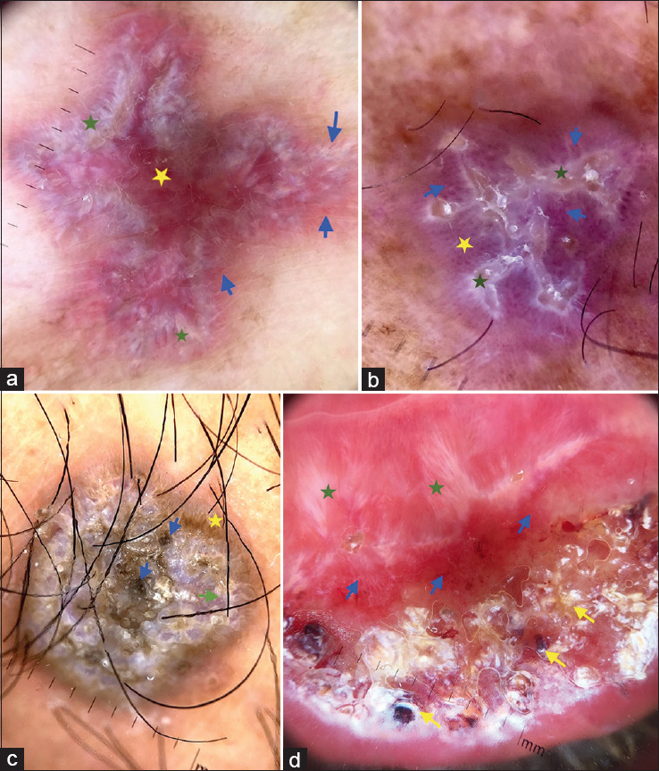
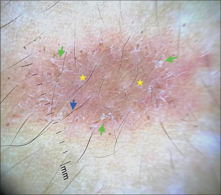
Dermatoscopy is a relevant diagnostic tool for inflammatory diseases of the skin that aids not only in diagnosis, but also in monitoring the response to treatment. The inflammatory diseases show dermoscopic patterns involving the vessels, scales, follicles, background hue, and special clues. This review aims to provide an overview on the use of dermoscopy in inflammatory dermatoses based on the available literature and the deviation from it in the skin of color (SOC) as there is paucity of literature in dermoscopy of inflammatory disorders in SOC. The dermatoscopic patterns in most of the inflammatory diseases in SOC are similar to that of white skin, with pigmentary changes being the prominent dermoscopic findings while vascular patterns and erythema being less evident.

摘要

皮肤镜检查是诊断皮肤炎症性疾病的一种重要工具,不仅有助于诊断,还能监测治疗反应。炎症性疾病呈现出涉及血管、鳞屑、毛囊、背景色调及特殊线索的皮肤镜特征。由于关于色素沉着皮肤炎症性疾病的皮肤镜检查文献较少,本综述旨在基于现有文献及色素沉着皮肤与该文献的差异,概述皮肤镜在炎症性皮肤病中的应用。色素沉着皮肤中大多数炎症性疾病的皮肤镜特征与白色皮肤相似,色素沉着改变是主要的皮肤镜表现,而血管形态和红斑则不那么明显。

https://cdn.ncbi.nlm.nih.gov/pmc/blobs/cc41/7982019/c2ed6d80234b/IDOJ-12-45-g022.jpg
https://cdn.ncbi.nlm.nih.gov/pmc/blobs/cc41/7982019/6321475069d2/IDOJ-12-45-g001.jpg
https://cdn.ncbi.nlm.nih.gov/pmc/blobs/cc41/7982019/a9c696aa7a4a/IDOJ-12-45-g002.jpg
https://cdn.ncbi.nlm.nih.gov/pmc/blobs/cc41/7982019/88590e342074/IDOJ-12-45-g003.jpg
https://cdn.ncbi.nlm.nih.gov/pmc/blobs/cc41/7982019/561b3a8d65e8/IDOJ-12-45-g004.jpg
https://cdn.ncbi.nlm.nih.gov/pmc/blobs/cc41/7982019/b34810c01a6e/IDOJ-12-45-g005.jpg
https://cdn.ncbi.nlm.nih.gov/pmc/blobs/cc41/7982019/d4269e952f65/IDOJ-12-45-g006.jpg
https://cdn.ncbi.nlm.nih.gov/pmc/blobs/cc41/7982019/72cbba415416/IDOJ-12-45-g007.jpg
https://cdn.ncbi.nlm.nih.gov/pmc/blobs/cc41/7982019/ad664019f6af/IDOJ-12-45-g008.jpg
https://cdn.ncbi.nlm.nih.gov/pmc/blobs/cc41/7982019/506efdef195d/IDOJ-12-45-g009.jpg
https://cdn.ncbi.nlm.nih.gov/pmc/blobs/cc41/7982019/1c09f69368e5/IDOJ-12-45-g010.jpg
https://cdn.ncbi.nlm.nih.gov/pmc/blobs/cc41/7982019/5ddc3623cf46/IDOJ-12-45-g011.jpg
https://cdn.ncbi.nlm.nih.gov/pmc/blobs/cc41/7982019/409841a5a743/IDOJ-12-45-g012.jpg
https://cdn.ncbi.nlm.nih.gov/pmc/blobs/cc41/7982019/6f772667f305/IDOJ-12-45-g013.jpg
https://cdn.ncbi.nlm.nih.gov/pmc/blobs/cc41/7982019/fc5ea1241c04/IDOJ-12-45-g014.jpg
https://cdn.ncbi.nlm.nih.gov/pmc/blobs/cc41/7982019/4aeac5c95cb0/IDOJ-12-45-g015.jpg
https://cdn.ncbi.nlm.nih.gov/pmc/blobs/cc41/7982019/215257c08f30/IDOJ-12-45-g016.jpg
https://cdn.ncbi.nlm.nih.gov/pmc/blobs/cc41/7982019/97ccbbe25253/IDOJ-12-45-g017.jpg
https://cdn.ncbi.nlm.nih.gov/pmc/blobs/cc41/7982019/9b6df1088a39/IDOJ-12-45-g018.jpg
https://cdn.ncbi.nlm.nih.gov/pmc/blobs/cc41/7982019/5f529d1e02c3/IDOJ-12-45-g019.jpg
https://cdn.ncbi.nlm.nih.gov/pmc/blobs/cc41/7982019/2af16a8eac8a/IDOJ-12-45-g020.jpg
https://cdn.ncbi.nlm.nih.gov/pmc/blobs/cc41/7982019/3cacbfad68d1/IDOJ-12-45-g021.jpg
https://cdn.ncbi.nlm.nih.gov/pmc/blobs/cc41/7982019/c2ed6d80234b/IDOJ-12-45-g022.jpg

相似文献

1
Dermatoscopy of Inflammatory Diseases in Skin of Color.有色人种皮肤炎症性疾病的皮肤镜检查
Indian Dermatol Online J. 2021 Jan 16;12(1):45-57. doi: 10.4103/idoj.IDOJ_613_20. eCollection 2021 Jan-Feb.
2
[Inflammoscopy: dermatoscopy for inflammatory, infiltrating and infectious dermatoses : Indication and standardization of dermatoscopic terminology].[炎症镜检:用于炎症性、浸润性和感染性皮肤病的皮肤镜检查:皮肤镜术语的指征与标准化]
Hautarzt. 2020 Aug;71(8):627-646. doi: 10.1007/s00105-020-04590-7.
3
Dermoscopy in general dermatology (non-neoplastic dermatoses) of skin of colour: a comparative retrospective study by the International Dermoscopy Society.皮肤色素性非肿瘤性皮肤病的皮肤镜检查:国际皮肤镜学会的一项比较回顾性研究。
Eur J Dermatol. 2020 Dec 1;30(6):688-698. doi: 10.1684/ejd.2020.3928.
4
Dermatoscopic Features of Pigmentary Diseases in Ethnic Skin.不同种族皮肤色素性疾病的皮肤镜特征
Indian Dermatol Online J. 2021 Jan 16;12(1):24-33. doi: 10.4103/idoj.IDOJ_561_20. eCollection 2021 Jan-Feb.
5
Dermoscopy of Inflammatory Dermatoses (Inflammoscopy): An Up-to-Date Overview.炎症性皮肤病的皮肤镜检查(炎症镜检):最新综述。
Dermatol Pract Concept. 2019 Jul 31;9(3):169-180. doi: 10.5826/dpc.0903a01. eCollection 2019 Jul.
6
Dermoscopy of Inflammatory Dermatoses (Inflammoscopy) in Skin of Color - A Systematic Review by the International Dermoscopy Society "Imaging in Skin of Color" Task Force.国际皮肤镜协会“有色人种皮肤成像”特别工作组对有色人种皮肤炎症性皮肤病的皮肤镜检查(炎症性皮肤镜检查)进行的系统评价
Dermatol Pract Concept. 2023 Oct 1;13(4 S1):e2023297S. doi: 10.5826/dpc.1304S1a297S.
7
Dermatoscopy of Cutaneous Lichen Planus - Attempt to Translate Metaphoric Terminology Into Descriptive Terminology.皮肤扁平苔藓的皮肤镜检查——尝试将隐喻性术语转化为描述性术语。
Dermatol Pract Concept. 2023 Jul 1;13(3):e2023174. doi: 10.5826/dpc.1303a174.
8
Dermatoscopy of Granulomatous Disorders.肉芽肿性疾病的皮肤镜检查
Dermatol Clin. 2018 Oct;36(4):369-375. doi: 10.1016/j.det.2018.05.004. Epub 2018 Aug 2.
9
Dermoscopy in the Diagnosis of Inflammatory Dermatoses: Systematic Review Findings Reported for Psoriasis, Lupus, and Lichen Planus.皮肤镜在炎症性皮肤病诊断中的应用:关于银屑病、狼疮和扁平苔藓的系统评价结果报告
J Clin Aesthet Dermatol. 2018 Apr;11(4):41-42. Epub 2018 Apr 1.
10
Dermoscopy of Common Inflammatory Disorders.常见炎症性疾病的皮肤镜检查
Dermatol Clin. 2018 Oct;36(4):359-368. doi: 10.1016/j.det.2018.05.003. Epub 2018 Jul 31.

引用本文的文献

1
Exploring Pediatric Dermatology in Skin of Color: Focus on Dermoscopy.探索有色人种儿童皮肤病学:聚焦皮肤镜检查。
Life (Basel). 2024 Dec 4;14(12):1604. doi: 10.3390/life14121604.
2
The mark of success: The role of vaccine-induced skin scar formation for BCG and smallpox vaccine-associated clinical benefits.成功的标志:疫苗诱导的皮肤瘢痕形成在卡介苗和天花疫苗相关临床获益中的作用。
Semin Immunopathol. 2024 Aug 26;46(5):13. doi: 10.1007/s00281-024-01022-9.
3
Dermoscopy of Inflammatory Dermatoses (Inflammoscopy) in Skin of Color - A Systematic Review by the International Dermoscopy Society "Imaging in Skin of Color" Task Force.

本文引用的文献

1
Dermoscopy of Urticaria Pigmentosa.色素性荨麻疹的皮肤镜检查
Indian Dermatol Online J. 2020 May 10;11(3):475-476. doi: 10.4103/idoj.IDOJ_438_18. eCollection 2020 May-Jun.
2
Dermoscopy of Localized Darier's Disease in Fitzpatrick Type IV Skin.IV型 Fitzpatrick皮肤中局限性 Darier病的皮肤镜检查
Indian Dermatol Online J. 2020 Mar 9;11(2):298-300. doi: 10.4103/idoj.IDOJ_412_18. eCollection 2020 Mar-Apr.
3
Mucoscopy of Discoid Lupus Erythematosus on Lower Lip.下唇盘状红斑狼疮的黏膜镜检查
国际皮肤镜协会“有色人种皮肤成像”特别工作组对有色人种皮肤炎症性皮肤病的皮肤镜检查(炎症性皮肤镜检查)进行的系统评价
Dermatol Pract Concept. 2023 Oct 1;13(4 S1):e2023297S. doi: 10.5826/dpc.1304S1a297S.
4
[Dermoscopy of nonneoplastic diseases in dark skin].[深色皮肤非肿瘤性疾病的皮肤镜检查]
Dermatologie (Heidelb). 2023 Apr;74(4):250-255. doi: 10.1007/s00105-023-05121-w. Epub 2023 Mar 1.
Indian Dermatol Online J. 2020 Mar 9;11(2):296-297. doi: 10.4103/idoj.IDOJ_348_18. eCollection 2020 Mar-Apr.
4
Dermoscopic features of various stages of lichen planus.扁平苔藓各阶段的皮肤镜特征。
Indian J Dermatol Venereol Leprol. 2020 Mar-Apr;86(2):191-194. doi: 10.4103/ijdvl.IJDVL_931_18.
5
Dermoscopy of Telangiectasia Macularis Eruptiva Perstans.持久性斑状毛细血管扩张性痣的皮肤镜检查
Indian Dermatol Online J. 2019 Apr 8;11(1):131-132. doi: 10.4103/idoj.IDOJ_312_18. eCollection 2020 Jan-Feb.
6
Dermoscopy Update: Review of Its Extradiagnostic and Expanding Indications and Future Prospects.皮肤镜最新进展:其诊断外及不断扩展的应用指征与未来前景综述
Dermatol Pract Concept. 2019 Oct 31;9(4):253-264. doi: 10.5826/dpc.0904a02. eCollection 2019 Oct.
7
Dermoscopy of Keratosis Pilaris.毛发角化病的皮肤镜检查
Indian Dermatol Online J. 2019 Aug 28;10(5):613-614. doi: 10.4103/idoj.IDOJ_279_18. eCollection 2019 Sep-Oct.
8
Role of Dermoscopy in the Diagnosis of Hypertrophic Lichen Planus and Prurigo Nodularis.皮肤镜在肥厚性扁平苔藓和结节性痒疹诊断中的作用
Indian J Dermatol. 2019 Sep-Oct;64(5):341-345. doi: 10.4103/ijd.IJD_123_18.
9
Dermoscopy of Inflammatory Dermatoses (Inflammoscopy): An Up-to-Date Overview.炎症性皮肤病的皮肤镜检查(炎症镜检):最新综述。
Dermatol Pract Concept. 2019 Jul 31;9(3):169-180. doi: 10.5826/dpc.0903a01. eCollection 2019 Jul.
10
Dermatoscopy of nonvenereal genital dermatoses: A brief review.非性病性生殖器皮肤病的皮肤镜检查:简要综述。
Indian J Sex Transm Dis AIDS. 2019 Jan-Jun;40(1):13-19. doi: 10.4103/ijstd.IJSTD_20_19.