Thomas Irene N, James Joseph Jenson, Bala Arishta, Mohan Saranya, Dogiparthi Sowmya, Shanmugam Nithya Priyadharshini
Dermatology, Shri Sathya Sai Medical College & Research Institute, Shri Balaji Vidyapeeth, Chennai, IND.
Dermatology, Shri Sathya Sai Medical College and Research Institute, Shri Balaji Vidyapeeth, Chennai, IND.
Cureus. 2023 Jun 11;15(6):e40271. doi: 10.7759/cureus.40271. eCollection 2023 Jun.
Hypopigmented patches in patients with skin of color are usually a cause of concern. Pityriasis alba is a common skin condition that causes visible patches of hypopigmentation in children and adolescents. In addition to the cosmetic impairment, parents are concerned about the diagnosis of vitiligo and leprosy which also cause hypopigmented patches and have negative social implications. Dermoscopy is a useful diagnostic aid that is acquiring prominence in diagnosing a variety of skin diseases. Few studies exist that validate the use of dermoscopy as an effective tool in the diagnosis of Pityriasis alba.
To evaluate the effectiveness of dermoscopy by correlating the clinical features of Pityriasis alba with dermoscopic images.
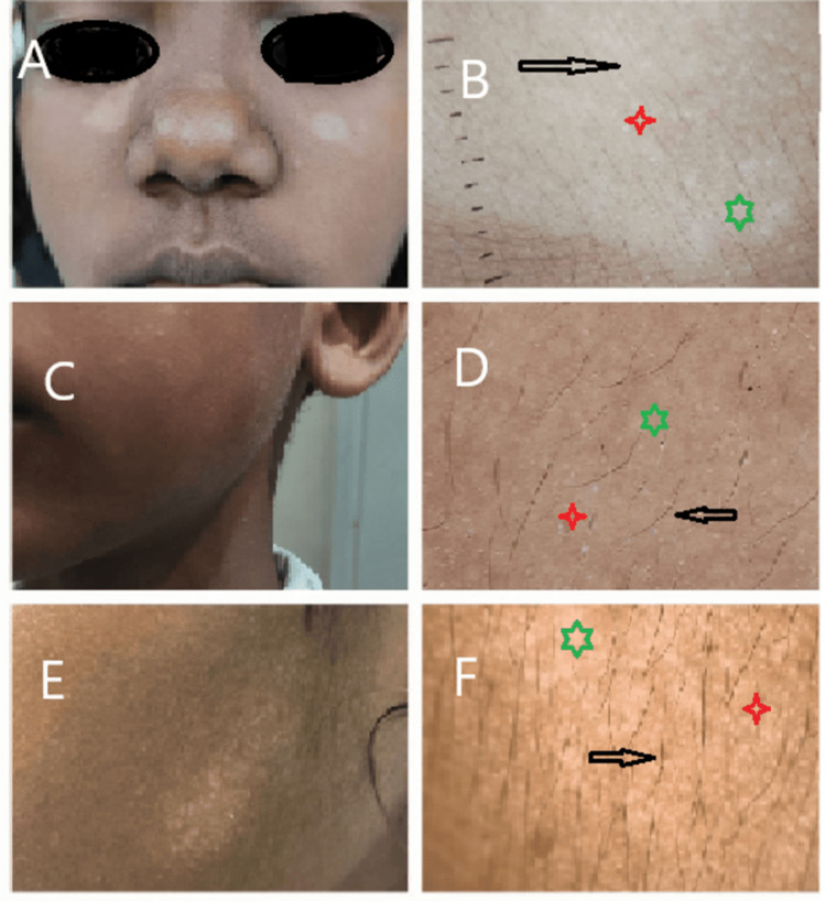
Hypopigmented patches in 16 patients that were clinically diagnosed as Pityriasis alba were examined with a DermLite DL200 Hybrid dermoscope (Dermlite, CA, USA). All the dermoscopic images were photographically recorded and the findings were noted and correlated with the clinical stages of the disease.
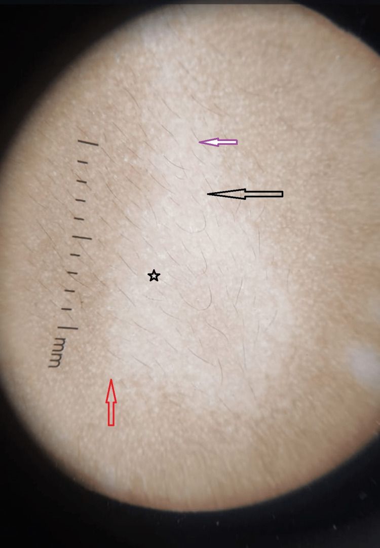
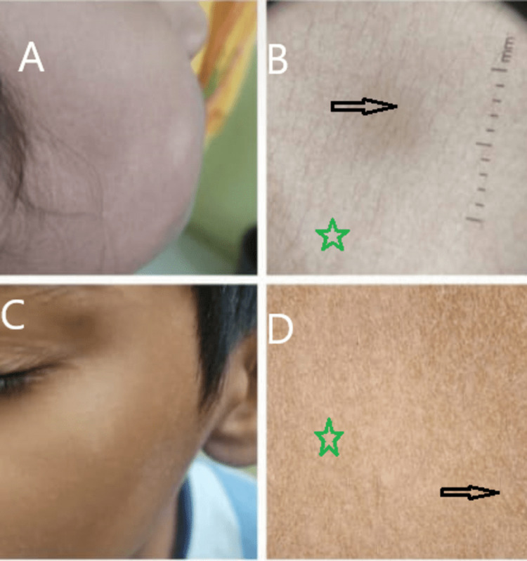
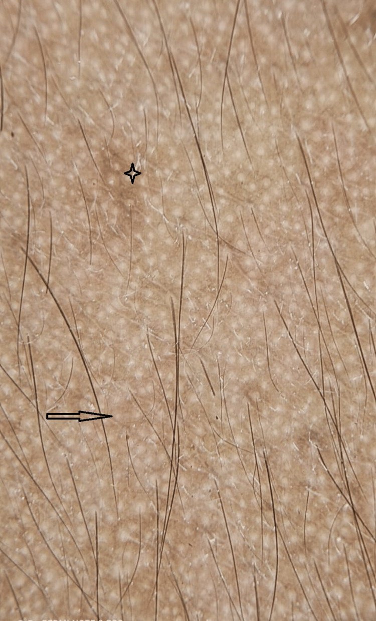
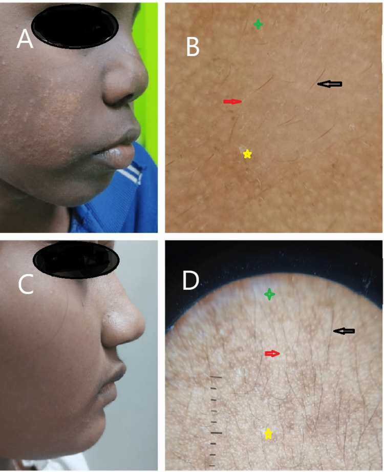
Out of the 40 patches examined in 16 patients, dermoscopic images of white structureless spots, scaling, indistinct borders and normally pigmented hairs were consistently present in all the patches to propose these as the four dermoscopic criteria for the diagnosis of Pityriasis alba. Areas of light brown pigmentation, 17 (42.5%), erythema, 3 (7.5%), and faint pigmented network,11 (27.5%) were the other features noted in some of the patches.
In an ethnic South Indian population where the skin color is predominantly brown, hypopigmented patches are visibly obvious and concerning. Pityriasis alba, Pityriasis versicolor, Vitiligo, Nevus depigmentosus, and Leprosy are the five common conditions seen among children of which Pityriasis alba is the most prevalent. Offering the right diagnosis is essential for the correct management as well as excluding more serious conditions such as leprosy and vitiligo. In this study, Dermoscopy provided a valuable diagnostic aid in achieving this objective.
有色人种患者的色素减退斑通常令人担忧。白色糠疹是一种常见的皮肤疾病,可导致儿童和青少年出现明显的色素减退斑。除了外观受损外,家长们还担心白癜风和麻风病的诊断,这两种疾病也会导致色素减退斑,并且具有负面的社会影响。皮肤镜检查是一种有用的诊断辅助手段,在诊断各种皮肤疾病方面正日益受到重视。很少有研究证实皮肤镜检查可作为诊断白色糠疹的有效工具。
通过将白色糠疹的临床特征与皮肤镜图像相关联,评估皮肤镜检查的有效性。
使用DermLite DL200 Hybrid皮肤镜(美国加利福尼亚州Dermlite公司)对16例临床诊断为白色糠疹的患者的色素减退斑进行检查。所有皮肤镜图像均拍照记录,并记录检查结果,使其与疾病的临床分期相关联。
在16例患者检查的40个斑块中,所有斑块均始终出现白色无结构斑点、鳞屑、边界不清和毛发色素正常的皮肤镜图像,据此提出将这些作为诊断白色糠疹的四个皮肤镜标准。在一些斑块中还发现了其他特征,如浅棕色色素沉着区域17处(42.5%)、红斑3处(7.5%)和淡色素网11处(27.5%)。
在皮肤颜色主要为棕色的印度南部人群中,色素减退斑明显可见且令人担忧。白色糠疹、花斑糠疹、白癜风、色素减退痣和麻风病是儿童中常见的五种疾病,其中白色糠疹最为普遍。提供正确的诊断对于正确治疗以及排除麻风病和白癜风等更严重的疾病至关重要。在本研究中,皮肤镜检查为实现这一目标提供了有价值的诊断辅助。