Department of Orthopedics, Orthopaedics Key Laboratory of Gansu Province, Lanzhou University Second Hospital, Lanzhou Gansu, China.
Channels (Austin). 2021 Dec;15(1):339-359. doi: 10.1080/19336950.2021.1903184.
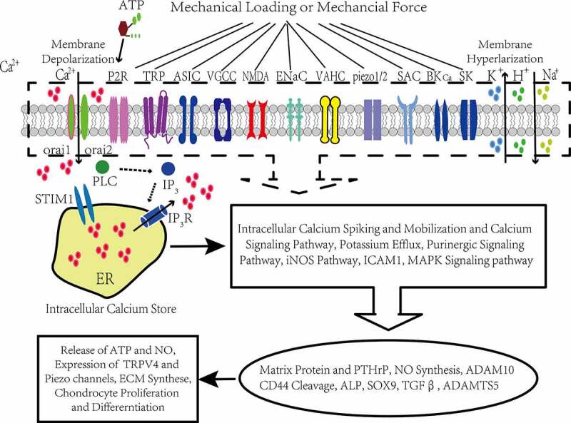
Articular cartilage consists of an extracellular matrix including many proteins as well as embedded chondrocytes. Articular cartilage formation and function are influenced by mechanical forces. Hind limb unloading or simulated microgravity causes articular cartilage loss, suggesting the importance of the healthy mechanical environment in articular cartilage homeostasis and implying a significant role of appropriate mechanical stimulation in articular cartilage degeneration. Mechanosensitive ion channels participate in regulating the metabolism of articular chondrocytes, including matrix protein production and extracellular matrix synthesis. Mechanical stimuli, including fluid shear stress, stretch, compression and cell swelling and decreased mechanical conditions (such as simulated microgravity) can alter the membrane potential and regulate the metabolism of articular chondrocytes via transmembrane ion channel-induced ionic fluxes. This process includes Ca influx and the resulting mobilization of Ca that is due to massive released Ca from stores, intracellular cation efflux and extracellular cation influx. This review brings together published information on mechanosensitive ion channels, such as stretch-activated channels (SACs), voltage-gated Ca channels (VGCCs), large conductance Ca-activated K channels (BK channels), Ca-activated K channels (SK channels), voltage-activated H channels (VAHCs), acid sensing ion channels (ASICs), transient receptor potential (TRP) family channels, and piezo1/2 channels. Data based on epithelial sodium channels (ENaCs), purinergic receptors and N-methyl-d-aspartate (NMDA) receptors are also included. These channels mediate mechanoelectrical physiological processes essential for converting physical force signals into biological signals. The primary channel-mediated effects and signaling pathways regulated by these mechanosensitive ion channels can influence the progression of osteoarthritis during the mechanosensory and mechanoadaptive process of articular chondrocytes.
关节软骨由细胞外基质组成,其中包括许多蛋白质和嵌入的软骨细胞。关节软骨的形成和功能受到机械力的影响。下肢去负荷或模拟微重力会导致关节软骨丢失,这表明健康的机械环境对关节软骨的稳态很重要,并暗示适当的机械刺激在关节软骨退变中起着重要作用。机械敏感离子通道参与调节关节软骨细胞的代谢,包括基质蛋白的产生和细胞外基质的合成。机械刺激,包括流体切应力、拉伸、压缩和细胞肿胀以及机械条件降低(如模拟微重力),可以通过跨膜离子通道诱导的离子流改变细胞膜电位并调节关节软骨细胞的代谢。这个过程包括 Ca 内流和 Ca 的大量释放,这是由于储存库中大量释放的 Ca,细胞内阳离子外流和细胞外阳离子内流所致。这篇综述汇集了关于机械敏感离子通道的已发表信息,如拉伸激活通道 (SACs)、电压门控 Ca 通道 (VGCCs)、大电导 Ca 激活的 K 通道 (BK 通道)、Ca 激活的 K 通道 (SK 通道)、电压激活的 H 通道 (VAHCs)、酸感应离子通道 (ASICs)、瞬时受体电位 (TRP) 家族通道和压电 1/2 通道。基于上皮钠通道 (ENaCs)、嘌呤能受体和 N-甲基-D-天冬氨酸 (NMDA) 受体的数据也包括在内。这些通道介导机械电生理过程,对于将物理力信号转化为生物信号至关重要。这些机械敏感离子通道调节的主要通道介导效应和信号通路可以影响关节软骨细胞的机械感觉和机械适应过程中骨关节炎的进展。