• 文献检索
  • 文档翻译
  • 深度研究
  • 学术资讯
  • Suppr Zotero 插件Zotero 插件
  • 邀请有礼
  • 套餐&价格
  • 历史记录
应用&插件
Suppr Zotero 插件Zotero 插件浏览器插件Mac 客户端Windows 客户端微信小程序
定价
高级版会员购买积分包购买API积分包
服务
文献检索文档翻译深度研究API 文档MCP 服务
关于我们
关于 Suppr公司介绍联系我们用户协议隐私条款
关注我们

Suppr 超能文献

核心技术专利:CN118964589B侵权必究
粤ICP备2023148730 号-1Suppr @ 2026

文献检索

告别复杂PubMed语法,用中文像聊天一样搜索,搜遍4000万医学文献。AI智能推荐,让科研检索更轻松。

立即免费搜索

文件翻译

保留排版,准确专业,支持PDF/Word/PPT等文件格式,支持 12+语言互译。

免费翻译文档

深度研究

AI帮你快速写综述,25分钟生成高质量综述,智能提取关键信息,辅助科研写作。

立即免费体验

用于肿瘤治疗的基于微流控的外泌体分离与检测

Towards Microfluidic-Based Exosome Isolation and Detection for Tumor Therapy.

作者信息

Wang Jie, Ma Peng, Kim Daniel H, Liu Bi-Feng, Demirci Utkan

机构信息

Canary Center at Stanford for Cancer Early Detection, Bio-Acoustic MEMS in Medicine (BAMM) Laboratory, Department of Radiology, School of Medicine Stanford University, Palo Alto, California 94304-5427, USA.

Canary Center at Stanford for Cancer Early Detection, Department of Radiology, Stanford University School of Medicine, Palo Alto, California 94305, USA.

出版信息

Nano Today. 2021 Apr;37. doi: 10.1016/j.nantod.2020.101066. Epub 2021 Jan 13.

DOI:10.1016/j.nantod.2020.101066
PMID:33777166
原文链接:https://pmc.ncbi.nlm.nih.gov/articles/PMC7990116/
Abstract

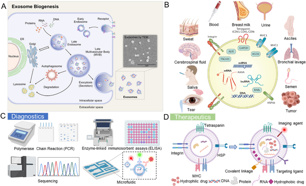
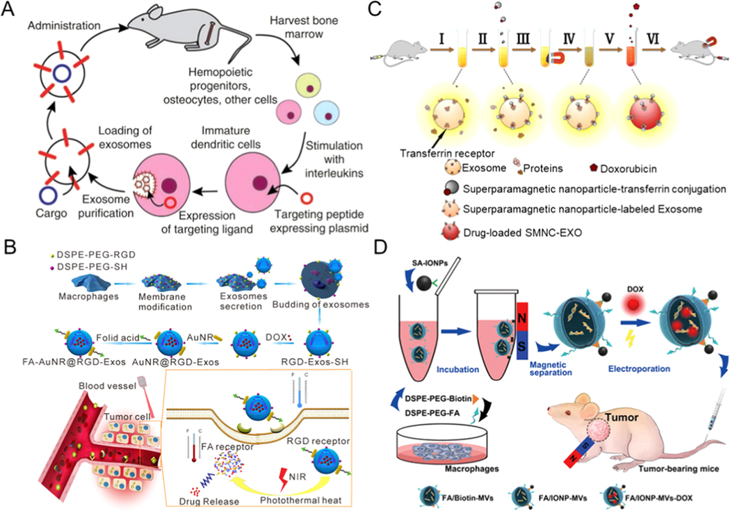
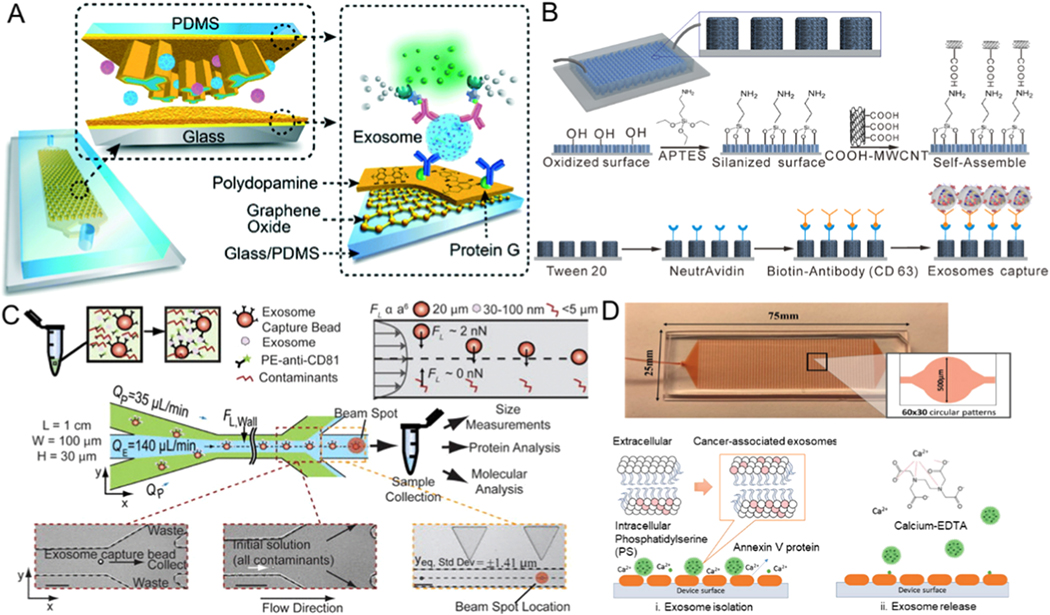
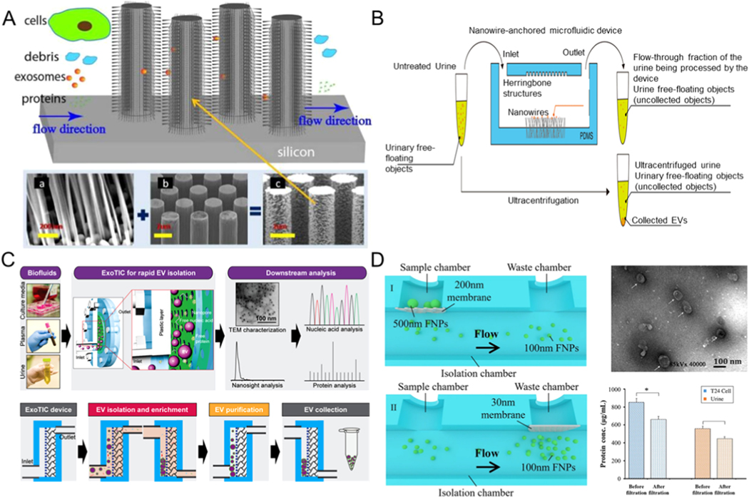
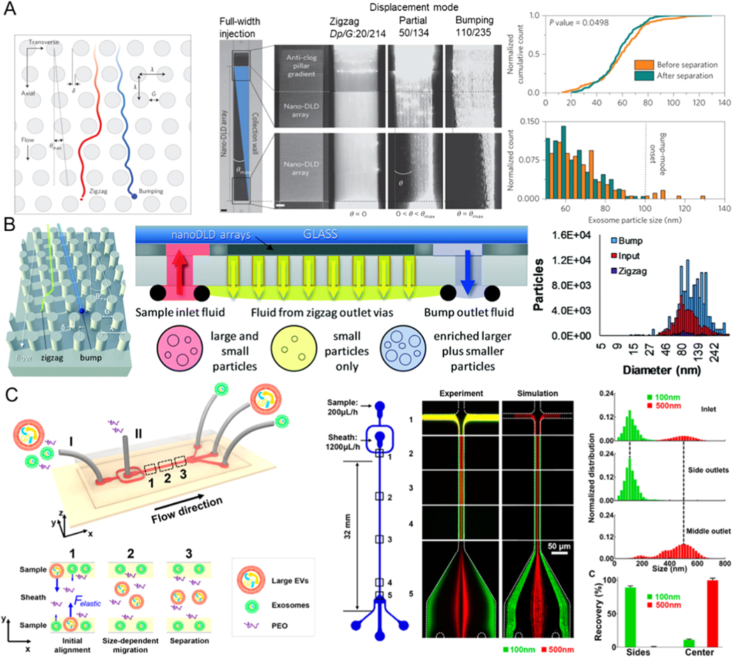
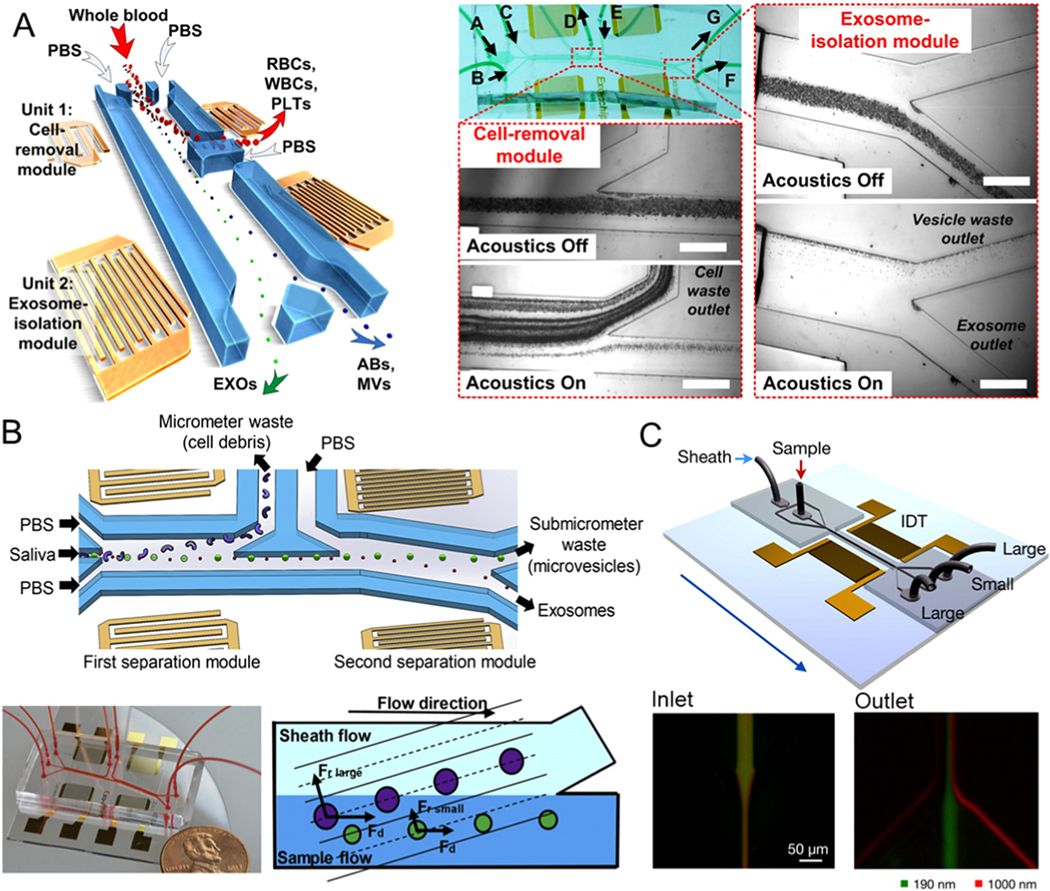
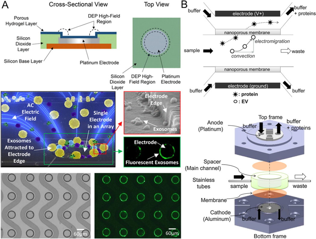
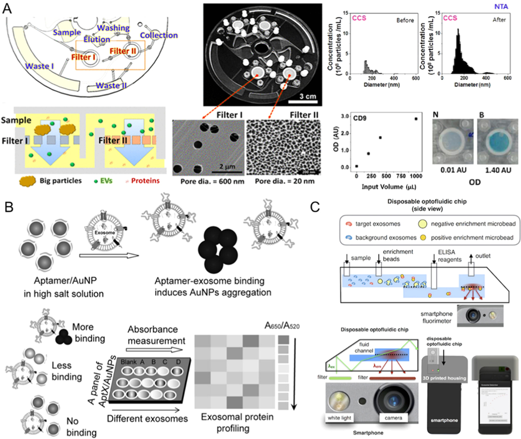
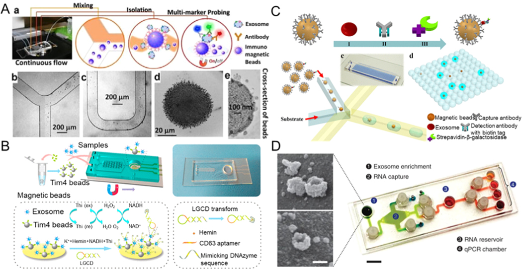
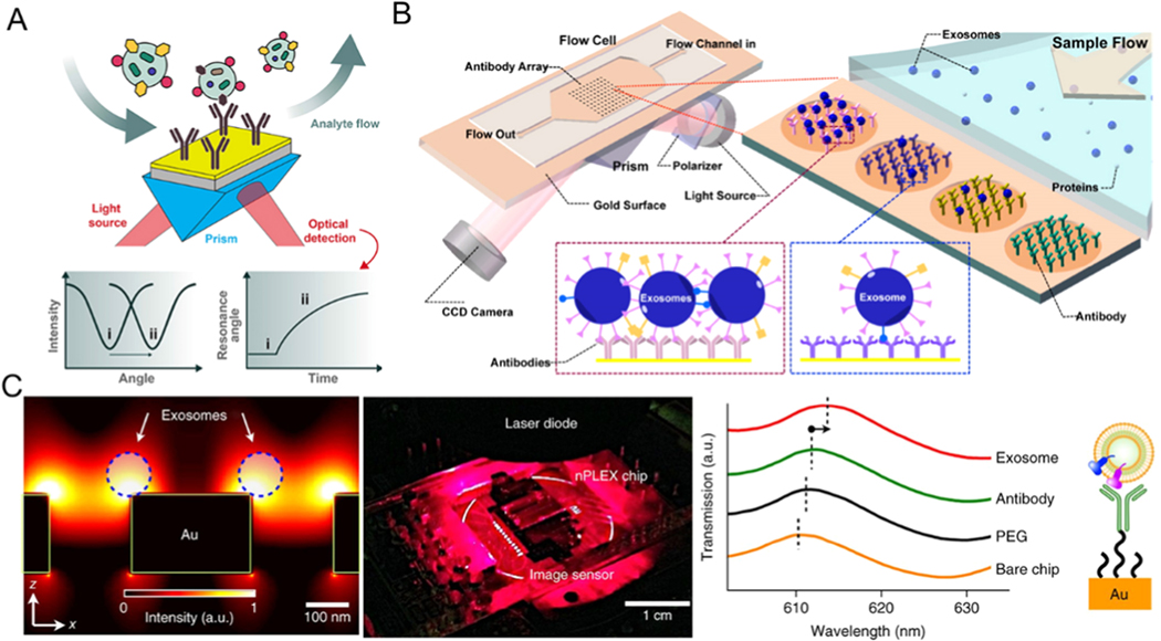
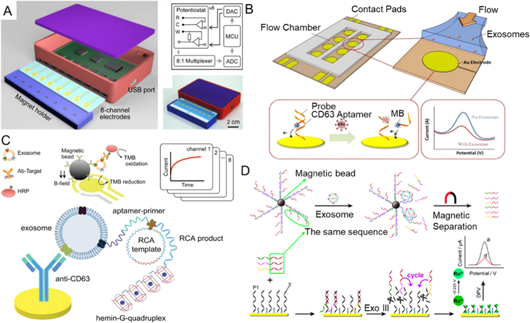
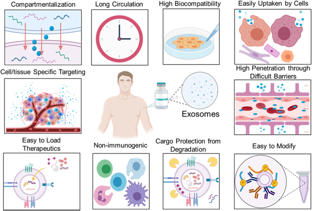
Exosomes are a class of cell-secreted, nano-sized extracellular vesicles with a bilayer membrane structure of 30-150 nm in diameter. Their discovery and application have brought breakthroughs in numerous areas, such as liquid biopsies, cancer biology, drug delivery, immunotherapy, tissue repair, and cardiovascular diseases. Isolation of exosomes is the first step in exosome-related research and its applications. Standard benchtop exosome separation and sensing techniques are tedious and challenging, as they require large sample volumes, multi-step operations that are complex and time-consuming, requiring cumbersome and expensive instruments. In contrast, microfluidic platforms have the potential to overcome some of these limitations, owing to their high-precision processing, ability to handle liquids at a microscale, and integrability with various functional units, such as mixers, actuators, reactors, separators, and sensors. These platforms can optimize the detection process on a single device, representing a robust and versatile technique for exosome separation and sensing to attain high purity and high recovery rates with a short processing time. Herein, we overview microfluidic strategies for exosome isolation based on their hydrodynamic properties, size filtration, acoustic fields, immunoaffinity, and dielectrophoretic properties. We focus especially on advances in label-free isolation of exosomes with active biological properties and intact morphological structures. Further, we introduce microfluidic techniques for the detection of exosomal proteins and RNAs with high sensitivity, high specificity, and low detection limits. We summarize the biomedical applications of exosome-mediated therapeutic delivery targeting cancer cells. To highlight the advantages of microfluidic platforms, conventional techniques are included for comparison. Future challenges and prospects of microfluidics towards exosome isolation applications are also discussed. Although the use of exosomes in clinical applications still faces biological, technical, regulatory, and market challenges, in the foreseeable future, recent developments in microfluidic technologies are expected to pave the way for tailoring exosome-related applications in precision medicine.

摘要

外泌体是一类细胞分泌的纳米级细胞外囊泡,具有直径为30 - 150 nm的双层膜结构。它们的发现和应用在众多领域带来了突破,如液体活检、癌症生物学、药物递送、免疫治疗、组织修复和心血管疾病等。外泌体的分离是外泌体相关研究及其应用的第一步。标准的台式外泌体分离和传感技术既繁琐又具有挑战性,因为它们需要大量样本体积、多步骤操作,这些操作复杂且耗时,还需要笨重且昂贵的仪器。相比之下,微流控平台有潜力克服其中一些限制,这得益于其高精度处理能力、在微尺度上处理液体的能力以及与各种功能单元(如混合器、致动器、反应器、分离器和传感器)的可集成性。这些平台可以在单个设备上优化检测过程,代表了一种强大且通用的外泌体分离和传感技术,能够在短处理时间内实现高纯度和高回收率。在此,我们概述基于外泌体流体动力学特性、尺寸过滤、声场、免疫亲和性和介电泳特性的外泌体分离微流控策略。我们特别关注具有活性生物学特性和完整形态结构的外泌体无标记分离方面的进展。此外,我们介绍用于高灵敏度、高特异性和低检测限检测外泌体蛋白质和RNA的微流控技术。我们总结了外泌体介导的靶向癌细胞治疗递送的生物医学应用。为突出微流控平台的优势,还纳入了传统技术进行比较。还讨论了微流控技术在外泌体分离应用方面未来面临的挑战和前景。尽管外泌体在临床应用中的使用仍面临生物学、技术、监管和市场方面的挑战,但在可预见的未来,微流控技术的最新进展有望为在精准医学中定制外泌体相关应用铺平道路。

https://cdn.ncbi.nlm.nih.gov/pmc/blobs/58d8/7990116/c65af4132d91/nihms-1658570-f0012.jpg
https://cdn.ncbi.nlm.nih.gov/pmc/blobs/58d8/7990116/eaeff539d392/nihms-1658570-f0001.jpg
https://cdn.ncbi.nlm.nih.gov/pmc/blobs/58d8/7990116/70f3808baebb/nihms-1658570-f0002.jpg
https://cdn.ncbi.nlm.nih.gov/pmc/blobs/58d8/7990116/a809f0dbec3a/nihms-1658570-f0003.jpg
https://cdn.ncbi.nlm.nih.gov/pmc/blobs/58d8/7990116/58fc0defce49/nihms-1658570-f0004.jpg
https://cdn.ncbi.nlm.nih.gov/pmc/blobs/58d8/7990116/7323bd8c5f94/nihms-1658570-f0005.jpg
https://cdn.ncbi.nlm.nih.gov/pmc/blobs/58d8/7990116/9541666ae0a6/nihms-1658570-f0006.jpg
https://cdn.ncbi.nlm.nih.gov/pmc/blobs/58d8/7990116/826be03a6ba9/nihms-1658570-f0007.jpg
https://cdn.ncbi.nlm.nih.gov/pmc/blobs/58d8/7990116/e32fc113cdc1/nihms-1658570-f0008.jpg
https://cdn.ncbi.nlm.nih.gov/pmc/blobs/58d8/7990116/4cd9a79f4472/nihms-1658570-f0009.jpg
https://cdn.ncbi.nlm.nih.gov/pmc/blobs/58d8/7990116/a21fee2ae865/nihms-1658570-f0010.jpg
https://cdn.ncbi.nlm.nih.gov/pmc/blobs/58d8/7990116/ca22f59db985/nihms-1658570-f0011.jpg
https://cdn.ncbi.nlm.nih.gov/pmc/blobs/58d8/7990116/c65af4132d91/nihms-1658570-f0012.jpg

相似文献

1
Towards Microfluidic-Based Exosome Isolation and Detection for Tumor Therapy.用于肿瘤治疗的基于微流控的外泌体分离与检测
Nano Today. 2021 Apr;37. doi: 10.1016/j.nantod.2020.101066. Epub 2021 Jan 13.
2
Exosome separation using microfluidic systems: size-based, immunoaffinity-based and dynamic methodologies.使用微流体系统分离外泌体:基于尺寸、基于免疫亲和和动态方法
Biotechnol J. 2017 Apr;12(4). doi: 10.1002/biot.201600699. Epub 2017 Feb 6.
3
[Microfluidic strategies for separation and analysis of circulating exosomes].[用于循环外泌体分离与分析的微流控策略]
Se Pu. 2021 Sep;39(9):968-980. doi: 10.3724/SP.J.1123.2021.07005.
4
Cancer Liquid Biopsy Using Integrated Microfluidic Exosome Analysis Platforms.癌症液体活检采用集成微流控外泌体分析平台。
Biotechnol J. 2020 May;15(5):e1900225. doi: 10.1002/biot.201900225. Epub 2020 Feb 20.
5
Review on Strategies and Technologies for Exosome Isolation and Purification.外泌体分离与纯化的策略与技术综述
Front Bioeng Biotechnol. 2022 Jan 5;9:811971. doi: 10.3389/fbioe.2021.811971. eCollection 2021.
6
Label-Free Isolation of Exosomes Using Microfluidic Technologies.微流控技术的无标记外泌体分离。
ACS Nano. 2021 Nov 23;15(11):17047-17079. doi: 10.1021/acsnano.1c03469. Epub 2021 Nov 1.
7
Exosome isolation using nanostructures and microfluidic devices.使用纳米结构和微流控装置进行外泌体分离。
Biomed Mater. 2021 Feb 17;16(2):022005. doi: 10.1088/1748-605X/abde70.
8
A Holistic Review of the State-of-the-Art Microfluidics for Exosome Separation: An Overview of the Current Status, Existing Obstacles, and Future Outlook.全面综述微流控技术在分离外泌体方面的最新进展:现状概述、现存障碍和未来展望。
Small. 2021 Jul;17(29):e2007174. doi: 10.1002/smll.202007174. Epub 2021 May 28.
9
The Yin and Yang of exosome isolation methods: conventional practice, microfluidics, and commercial kits.外泌体分离方法的阴阳:传统方法、微流控技术及商业试剂盒
Biotechnol Adv. 2022 Jan-Feb;54:107814. doi: 10.1016/j.biotechadv.2021.107814. Epub 2021 Aug 11.
10
Progress in Microfluidics-Based Exosome Separation and Detection Technologies for Diagnostic Applications.基于微流控的外泌体分离和检测技术在诊断应用中的进展。
Small. 2020 Mar;16(9):e1903916. doi: 10.1002/smll.201903916. Epub 2019 Oct 30.

引用本文的文献

1
Mesenchymal stem cell-derived exosomes as a potential therapeutic strategy for ferroptosis.间充质干细胞衍生的外泌体作为铁死亡的一种潜在治疗策略。
Stem Cell Res Ther. 2025 Jul 15;16(1):368. doi: 10.1186/s13287-025-04511-2.
2
Adipose-derived stem cell exosomes: mechanisms and therapeutic potentials in wound healing.脂肪来源干细胞外泌体:伤口愈合中的作用机制及治疗潜力
Biomark Res. 2025 Jun 20;13(1):88. doi: 10.1186/s40364-025-00801-2.
3
Isolation methods of exosomes derived from dental stem cells.从牙源性干细胞中提取外泌体的分离方法。

本文引用的文献

1
Atlas of Exosomal microRNAs Secreted From Human iPSC-Derived Cardiac Cell Types.人诱导多能干细胞衍生心肌细胞类型分泌的外泌体微小RNA图谱
Circulation. 2020 Nov 3;142(18):1794-1796. doi: 10.1161/CIRCULATIONAHA.120.048364. Epub 2020 Nov 2.
2
Extracellular Vesicle and Particle Biomarkers Define Multiple Human Cancers.细胞外囊泡和颗粒生物标志物可定义多种人类癌症。
Cell. 2020 Aug 20;182(4):1044-1061.e18. doi: 10.1016/j.cell.2020.07.009. Epub 2020 Aug 13.
3
Entangled Nanoplasmonic Cavities for Estimating Thickness of Surface-Adsorbed Layers.
Int J Oral Sci. 2025 Jun 16;17(1):50. doi: 10.1038/s41368-025-00370-y.
4
Engineered brain-targeting exosome for reprogramming immunosuppressive microenvironment of glioblastoma.工程化脑靶向外泌体用于重编程胶质母细胞瘤的免疫抑制微环境
Exploration (Beijing). 2024 Jun 26;5(2):20240039. doi: 10.1002/EXP.20240039. eCollection 2025 Apr.
5
Exosome-Based Therapeutics in Dermatology.基于外泌体的皮肤病治疗方法
Biomater Res. 2025 May 9;29:0148. doi: 10.34133/bmr.0148. eCollection 2025.
6
[Exosome separation and analysis based on microfluidics technology and its clinical applications].基于微流控技术的外泌体分离与分析及其临床应用
Se Pu. 2025 May;43(5):455-471. doi: 10.3724/SP.J.1123.2024.10032.
7
The Roles of Exosomes in Anti-Cancer Drugs.外泌体在抗癌药物中的作用。
Cancer Med. 2025 May;14(9):e70897. doi: 10.1002/cam4.70897.
8
Small extracellular vesicle-based one-step high-throughput microfluidic platform for epithelial ovarian cancer diagnosis.用于上皮性卵巢癌诊断的基于小细胞外囊泡的一步式高通量微流控平台。
J Nanobiotechnology. 2025 Apr 7;23(1):278. doi: 10.1186/s12951-025-03348-4.
9
Exosome isolation and characterization for advanced diagnostic and therapeutic applications.用于先进诊断和治疗应用的外泌体分离与表征。
Mater Today Bio. 2025 Feb 25;31:101613. doi: 10.1016/j.mtbio.2025.101613. eCollection 2025 Apr.
10
Proteomics of Extracellular Vesicles: Recent Updates, Challenges and Limitations.细胞外囊泡蛋白质组学:最新进展、挑战与局限
Proteomes. 2025 Mar 4;13(1):12. doi: 10.3390/proteomes13010012.
用于估计表面吸附层厚度的纠缠纳米等离子体腔。
ACS Nano. 2020 Jul 28;14(7):8518-8527. doi: 10.1021/acsnano.0c02797. Epub 2020 Jul 8.
4
Clinical utility of the exosome based ExoDx Prostate(IntelliScore) EPI test in men presenting for initial Biopsy with a PSA 2-10 ng/mL.基于外泌体的 ExoDx Prostate(IntelliScore) EPI 检测在 PSA 2-10ng/mL 初始活检男性中的临床应用。
Prostate Cancer Prostatic Dis. 2020 Dec;23(4):607-614. doi: 10.1038/s41391-020-0237-z. Epub 2020 May 7.
5
Progress, opportunity, and perspective on exosome isolation - efforts for efficient exosome-based theranostics.外泌体分离的进展、机遇和展望——基于外泌体的高效治疗学的努力。
Theranostics. 2020 Feb 19;10(8):3684-3707. doi: 10.7150/thno.41580. eCollection 2020.
6
Thermophoretic Detection of Exosomal microRNAs by Nanoflares.纳米耀斑实现外泌体 microRNAs 的热泳检测
J Am Chem Soc. 2020 Mar 18;142(11):4996-5001. doi: 10.1021/jacs.9b13960. Epub 2020 Mar 6.
7
Engineering of Exosomes to Target Cancer Metastasis.工程化外泌体以靶向癌症转移
Cell Mol Bioeng. 2019 Dec 23;13(1):1-16. doi: 10.1007/s12195-019-00607-x. eCollection 2020 Feb.
8
Acoustofluidic Salivary Exosome Isolation: A Liquid Biopsy Compatible Approach for Human Papillomavirus-Associated Oropharyngeal Cancer Detection.声流唾液外泌体分离:一种适用于人乳头瘤病毒相关口咽癌检测的液体活检方法。
J Mol Diagn. 2020 Jan;22(1):50-59. doi: 10.1016/j.jmoldx.2019.08.004.
9
Exosome redux.外泌体再现。
Nat Biotechnol. 2019 Dec;37(12):1395-1400. doi: 10.1038/s41587-019-0326-5.
10
Electrokinetically Driven Exosome Separation and Concentration Using Dielectrophoretic-Enhanced PDMS-Based Microfluidics.基于电介质电泳增强的 PDMS 微流控的电动驱动外泌体分离和浓缩。
Anal Chem. 2019 Dec 3;91(23):14975-14982. doi: 10.1021/acs.analchem.9b03448. Epub 2019 Nov 18.