• 文献检索
  • 文档翻译
  • 深度研究
  • 学术资讯
  • Suppr Zotero 插件Zotero 插件
  • 邀请有礼
  • 套餐&价格
  • 历史记录
应用&插件
Suppr Zotero 插件Zotero 插件浏览器插件Mac 客户端Windows 客户端微信小程序
定价
高级版会员购买积分包购买API积分包
服务
文献检索文档翻译深度研究API 文档MCP 服务
关于我们
关于 Suppr公司介绍联系我们用户协议隐私条款
关注我们

Suppr 超能文献

核心技术专利:CN118964589B侵权必究
粤ICP备2023148730 号-1Suppr @ 2026

文献检索

告别复杂PubMed语法,用中文像聊天一样搜索,搜遍4000万医学文献。AI智能推荐,让科研检索更轻松。

立即免费搜索

文件翻译

保留排版,准确专业,支持PDF/Word/PPT等文件格式,支持 12+语言互译。

免费翻译文档

深度研究

AI帮你快速写综述,25分钟生成高质量综述,智能提取关键信息,辅助科研写作。

立即免费体验

胸部CT上新冠肺炎肺炎的定量负担可预测不良结局:一项前瞻性国际登记处的事后分析

Quantitative Burden of COVID-19 Pneumonia on Chest CT Predicts Adverse Outcomes: A Post-Hoc Analysis of a Prospective International Registry.

作者信息

Grodecki Kajetan, Lin Andrew, Cadet Sebastien, McElhinney Priscilla A, Razipour Aryabod, Chan Cato, Pressman Barry, Julien Peter, Maurovich-Horvat Pal, Gaibazzi Nicola, Thakur Udit, Mancini Elisabetta, Agalbato Cecilia, Menè Roberto, Parati Gianfranco, Cernigliaro Franco, Nerlekar Nitesh, Torlasco Camilla, Pontone Gianluca, Slomka Piotr J, Dey Damini

机构信息

Biomedical Imaging Research Institute, Cedars-Sinai Medical Center, Los Angeles, CA, USA (K.G., A.L., P.A.M., A.R., D.D.); Monash Health, Melbourne, Australia (A.L., U.T., N.N.); Department of Imaging, Cedars-Sinai Medical Center (S.C., C.C., B.P., P.J.); Department of Radiology, Semmelweis University, Budapest, Hungary (P.M.); Cardiology, Azienda Ospedaliero-Universitaria di Parma, Parma Italy (N.G.); Centro Cardiologico Monzino IRCCS, University of Milan, Italy (E.M., C.A., G.P.); Department of Cardiovascular, Neural and Metabolic Sciences, IRCCS Istituto Auxologico Italiano, Milan, Italy; Department of Medicine and Surgery, University of Milano-Bicocca, Italy (R.M., G.P., F.C., C.T.); Artificial Intelligence in Medicine Program, Cedars-Sinai Medical Center, Los Angeles, CA, USA (P.J.S.).

出版信息

Radiol Cardiothorac Imaging. 2020 Oct 1;2(5):e200389. doi: 10.1148/ryct.2020200389. eCollection 2020 Oct.

DOI:10.1148/ryct.2020200389
PMID:33778629
原文链接:https://pmc.ncbi.nlm.nih.gov/articles/PMC7605078/
Abstract

PURPOSE

To examine the independent and incremental value of CT-derived quantitative burden and attenuation of COVID-19 pneumonia for the prediction of clinical deterioration or death.

METHODS

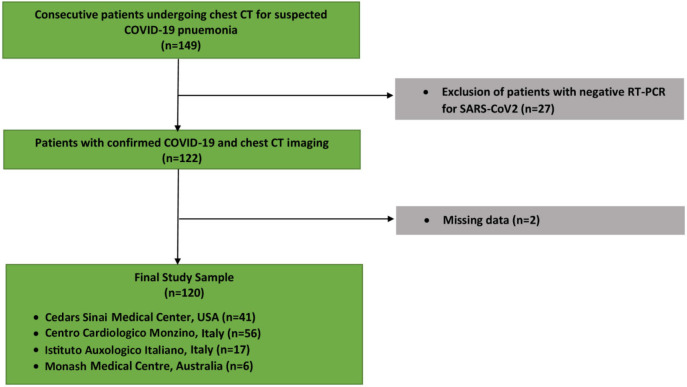
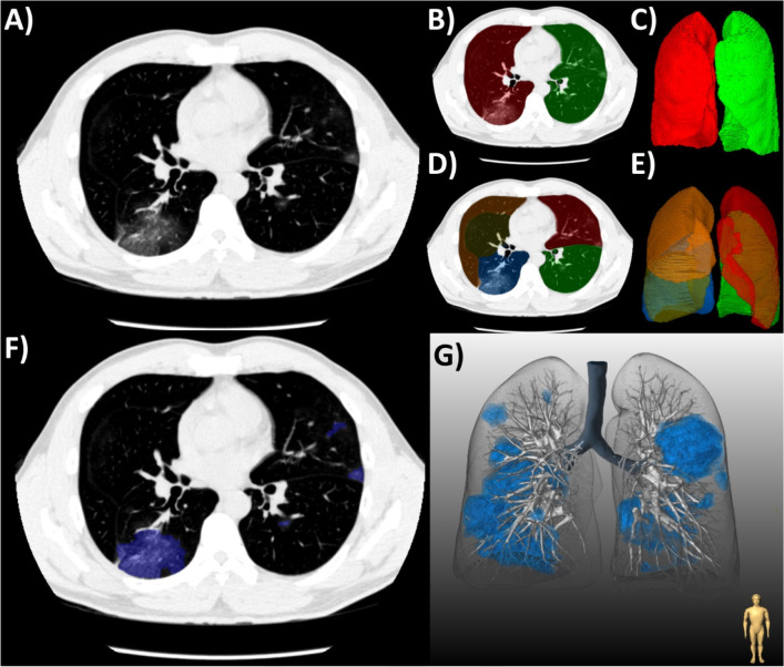
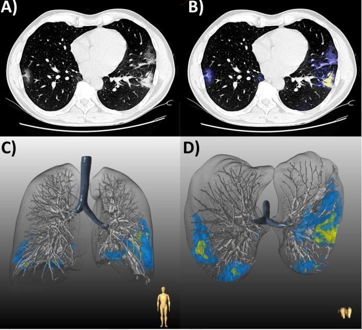
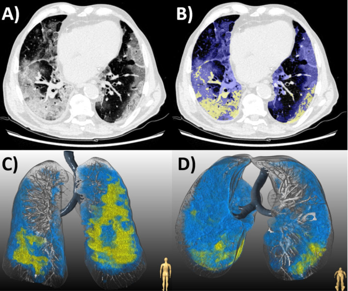
This was a retrospective analysis of a prospective international registry of consecutive patients with laboratory-confirmed COVID-19 and chest CT imaging, admitted to four centers between January 10 and May 6, 2020. Total burden (expressed as a percentage) and mean attenuation of ground glass opacities (GGO) and consolidation were quantified from CT using semi-automated research software. The primary outcome was clinical deterioration (intensive care unit admission, invasive mechanical ventilation, or vasopressor therapy) or in-hospital death. Logistic regression was performed to assess the predictive value of clinical and CT parameters for the primary outcome.

RESULTS

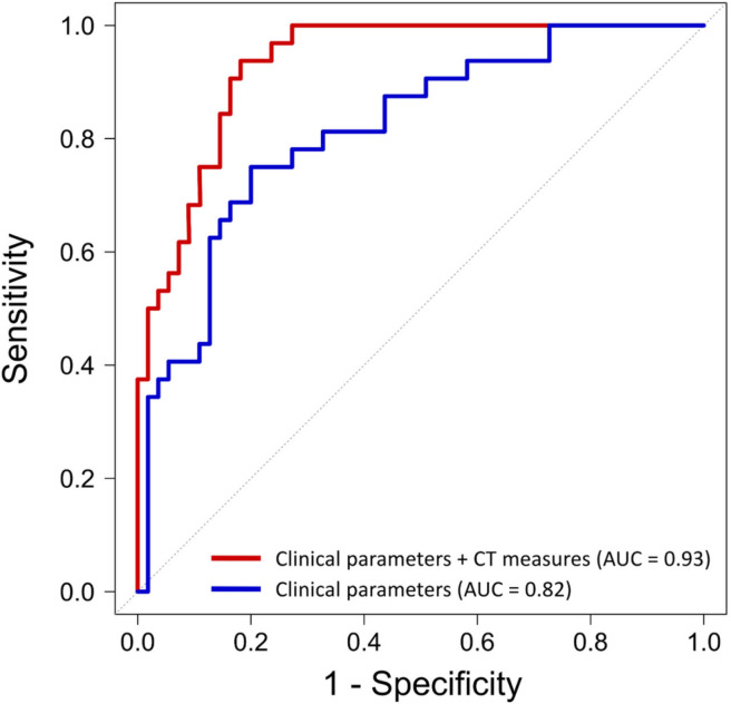
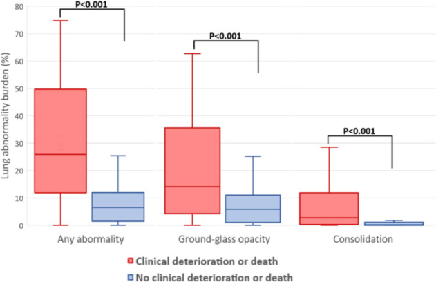
The final population comprised 120 patients (mean age 64 ± 16 years, 78 men), of whom 39 (32.5%) experienced clinical deterioration or death. In multivariable regression of clinical and CT parameters, consolidation burden (odds ratio [OR], 3.4; 95% confidence interval [CI]: 1.7, 6.9 per doubling; = .001) and increasing GGO attenuation (OR, 3.2; 95% CI: 1.3, 8.3 per standard deviation, = .02) were independent predictors of deterioration or death; as was C-reactive protein (OR, 2.1; 95% CI: 1.3, 3.4 per doubling; = .004), history of heart failure (OR 1.3; 95% CI: 1.1, 1.6, = .01), and chronic lung disease (OR, 1.3; 95% CI: 1.0, 1.6; = .02). Quantitative CT measures added incremental predictive value beyond a model with only clinical parameters (area under the curve, 0.93 vs 0.82, = .006). The optimal prognostic cutoffs for burden of COVID-19 pneumonia as determined by Youden's index were consolidation of greater than or equal to 1.8% and GGO of greater than or equal to 13.5%.

CONCLUSIONS

Quantitative burden of consolidation or GGO on chest CT independently predict clinical deterioration or death in patients with COVID-19 pneumonia. CT-derived measures have incremental prognostic value over and above clinical parameters, and may be useful for risk stratifying patients with COVID-19.

摘要

目的

探讨CT衍生的新型冠状病毒肺炎定量负担和衰减对预测临床恶化或死亡的独立及增量价值。

方法

这是一项对2020年1月10日至5月6日期间入住四个中心的连续实验室确诊新型冠状病毒肺炎且有胸部CT影像的患者进行的前瞻性国际登记研究的回顾性分析。使用半自动研究软件从CT中量化总负担(以百分比表示)以及磨玻璃影(GGO)和实变的平均衰减。主要结局为临床恶化(入住重症监护病房、有创机械通气或血管活性药物治疗)或院内死亡。进行逻辑回归以评估临床和CT参数对主要结局的预测价值。

结果

最终纳入120例患者(平均年龄64±16岁,男性78例),其中39例(32.5%)出现临床恶化或死亡。在临床和CT参数的多变量回归中,实变负担(比值比[OR],3.4;95%置信区间[CI]:每增加一倍为1.7,6.9;P = 0.001)和GGO衰减增加(OR,3.2;95%CI:每标准差为1.3,8.3;P = 0.02)是恶化或死亡的独立预测因素;C反应蛋白(OR,2.1;95%CI:每增加一倍为1.3,3.4;P = 0.004)、心力衰竭病史(OR 1.3;95%CI:1.1,1.6;P = 0.01)和慢性肺病(OR,1.3;95%CI:1.0,1.6;P = 0.02)也是如此。定量CT测量在仅包含临床参数的模型之外增加了增量预测价值(曲线下面积,0.93对vs0.82;P = 0.006)。根据约登指数确定的新型冠状病毒肺炎负担的最佳预后临界值为实变大于或等于1.8%和GGO大于或等于13.5%。

结论

胸部CT上实变或GGO的定量负担可独立预测新型冠状病毒肺炎患者的临床恶化或死亡。CT衍生的测量在临床参数之外具有增量预后价值,可能有助于对新型冠状病毒肺炎患者进行风险分层。

https://cdn.ncbi.nlm.nih.gov/pmc/blobs/d7b9/7977795/6a84730ba17f/ryct.2020200389.fig6.jpg
https://cdn.ncbi.nlm.nih.gov/pmc/blobs/d7b9/7977795/28d56cf5b91d/ryct.2020200389.fig1.jpg
https://cdn.ncbi.nlm.nih.gov/pmc/blobs/d7b9/7977795/87508b86b615/ryct.2020200389.fig2.jpg
https://cdn.ncbi.nlm.nih.gov/pmc/blobs/d7b9/7977795/37da74896d95/ryct.2020200389.fig3.jpg
https://cdn.ncbi.nlm.nih.gov/pmc/blobs/d7b9/7977795/4c9527e018fd/ryct.2020200389.fig4.jpg
https://cdn.ncbi.nlm.nih.gov/pmc/blobs/d7b9/7977795/c5680a64fb99/ryct.2020200389.fig5.jpg
https://cdn.ncbi.nlm.nih.gov/pmc/blobs/d7b9/7977795/6a84730ba17f/ryct.2020200389.fig6.jpg

相似文献

1
Quantitative Burden of COVID-19 Pneumonia on Chest CT Predicts Adverse Outcomes: A Post-Hoc Analysis of a Prospective International Registry.胸部CT上新冠肺炎肺炎的定量负担可预测不良结局:一项前瞻性国际登记处的事后分析
Radiol Cardiothorac Imaging. 2020 Oct 1;2(5):e200389. doi: 10.1148/ryct.2020200389. eCollection 2020 Oct.
2
Epicardial adipose tissue is associated with extent of pneumonia and adverse outcomes in patients with COVID-19.心外膜脂肪组织与 COVID-19 患者肺炎的严重程度和不良结局相关。
Metabolism. 2021 Feb;115:154436. doi: 10.1016/j.metabol.2020.154436. Epub 2020 Nov 19.
3
Automated AI-Driven CT Quantification of Lung Disease Predicts Adverse Outcomes in Patients Hospitalized for COVID-19 Pneumonia.基于人工智能的肺部疾病CT自动定量分析可预测COVID-19肺炎住院患者的不良预后。
Diagnostics (Basel). 2021 May 14;11(5):878. doi: 10.3390/diagnostics11050878.
4
Artificial intelligence-assisted quantification of COVID-19 pneumonia burden from computed tomography improves prediction of adverse outcomes over visual scoring systems.人工智能辅助 CT 对 COVID-19 肺炎负担的定量评估可提高对不良结局的预测能力,优于视觉评分系统。
Br J Radiol. 2023 Sep;96(1149):20220180. doi: 10.1259/bjr.20220180. Epub 2023 Jun 27.
5
[Spatial and temporal distribution and predictive value of chest CT scoring in patients with COVID-19].新型冠状病毒肺炎患者胸部CT评分的时空分布及预测价值
Zhonghua Jie He He Hu Xi Za Zhi. 2021 Mar 12;44(3):230-236. doi: 10.3760/cma.j.cn112147-20200522-00626.
6
Clinical and laboratory data, radiological structured report findings and quantitative evaluation of lung involvement on baseline chest CT in COVID-19 patients to predict prognosis.对 COVID-19 患者基线胸部 CT 上的临床和实验室数据、放射学结构化报告结果以及肺部受累的定量评估,以预测其预后。
Radiol Med. 2021 Jan;126(1):29-39. doi: 10.1007/s11547-020-01293-w. Epub 2020 Oct 12.
7
Value of quantitative analysis in lung computed tomography in patients severely ill with COVID-19.COVID-19 重症患者肺部计算机断层扫描的定量分析价值。
PLoS One. 2021 May 20;16(5):e0251946. doi: 10.1371/journal.pone.0251946. eCollection 2021.
8
CT Quantitative Analysis and Its Relationship with Clinical Features for Assessing the Severity of Patients with COVID-19.CT 定量分析及其与临床特征的关系,用于评估 COVID-19 患者的严重程度。
Korean J Radiol. 2020 Jul;21(7):859-868. doi: 10.3348/kjr.2020.0293.
9
Well-aerated Lung on Admitting Chest CT to Predict Adverse Outcome in COVID-19 Pneumonia.胸部 CT 显示充气良好的肺可预测 COVID-19 肺炎的不良结局。
Radiology. 2020 Aug;296(2):E86-E96. doi: 10.1148/radiol.2020201433. Epub 2020 Apr 17.
10
Comparison of chest radiography and chest CT for evaluation of pediatric COVID-19 pneumonia: Does CT add diagnostic value?比较胸部 X 线摄影和胸部 CT 对儿童 COVID-19 肺炎的评估:CT 是否增加诊断价值?
Pediatr Pulmonol. 2021 Jun;56(6):1409-1418. doi: 10.1002/ppul.25313. Epub 2021 Feb 25.

引用本文的文献

1
Artificial intelligence-based analysis of the spatial distribution of abnormal computed tomography patterns in SARS-CoV-2 pneumonia: association with disease severity.基于人工智能的 SARS-CoV-2 肺炎异常计算机断层扫描模式的空间分布分析:与疾病严重程度的关系。
Respir Res. 2024 Jan 10;25(1):24. doi: 10.1186/s12931-024-02673-w.
2
Patients Hospitalized for COVID-19 in the Periods of Delta and Omicron Variant Dominance in Greece: Determinants of Severity and Mortality.希腊在德尔塔和奥密克戎变异株主导时期因新冠病毒病住院的患者:严重程度和死亡率的决定因素
J Clin Med. 2023 Sep 11;12(18):5904. doi: 10.3390/jcm12185904.
3
Artificial intelligence-assisted quantification of COVID-19 pneumonia burden from computed tomography improves prediction of adverse outcomes over visual scoring systems.

本文引用的文献

1
Radiological Society of North America Expert Consensus Document on Reporting Chest CT Findings Related to COVID-19: Endorsed by the Society of Thoracic Radiology, the American College of Radiology, and RSNA.北美放射学会关于报告与COVID-19相关的胸部CT检查结果的专家共识文件:得到了胸腔放射学会、美国放射学会和北美放射学会的认可。
Radiol Cardiothorac Imaging. 2020 Mar 25;2(2):e200152. doi: 10.1148/ryct.2020200152. eCollection 2020 Apr.
2
CT Features and Short-term Prognosis of COVID-19 Pneumonia: A Single-Center Study from Kashan, Iran.新型冠状病毒肺炎的CT特征及短期预后:来自伊朗卡尚的一项单中心研究
Radiol Cardiothorac Imaging. 2020 Apr 20;2(2):e200130. doi: 10.1148/ryct.2020200130. eCollection 2020 Apr.
3
人工智能辅助 CT 对 COVID-19 肺炎负担的定量评估可提高对不良结局的预测能力,优于视觉评分系统。
Br J Radiol. 2023 Sep;96(1149):20220180. doi: 10.1259/bjr.20220180. Epub 2023 Jun 27.
4
A deep learning-based application for COVID-19 diagnosis on CT: The Imaging COVID-19 AI initiative.基于深度学习的 CT 新冠肺炎诊断应用:影像 COVID-19 AI 计划。
PLoS One. 2023 May 2;18(5):e0285121. doi: 10.1371/journal.pone.0285121. eCollection 2023.
5
Is the generalizability of a developed artificial intelligence algorithm for COVID-19 on chest CT sufficient for clinical use? Results from the International Consortium for COVID-19 Imaging AI (ICOVAI).开发用于 COVID-19 胸部 CT 的人工智能算法的可推广性是否足以用于临床?来自 COVID-19 成像人工智能国际联合会(ICOVAI)的结果。
Eur Radiol. 2023 Jun;33(6):4249-4258. doi: 10.1007/s00330-022-09303-3. Epub 2023 Jan 18.
6
Radiomorphological signs and clinical severity of SARS-CoV-2 lineage B.1.1.7.严重急性呼吸综合征冠状病毒2(SARS-CoV-2)B.1.1.7谱系的放射形态学特征及临床严重程度
BJR Open. 2022 May 6;4(1):20220016. doi: 10.1259/bjro.20220016. eCollection 2022.
7
CT Examinations for COVID-19: A Systematic Review of Protocols, Radiation Dose, and Numbers Needed to Diagnose and Predict.用于新型冠状病毒肺炎的CT检查:对方案、辐射剂量以及诊断和预测所需数量的系统评价
Taehan Yongsang Uihakhoe Chi. 2021 Nov;82(6):1505-1523. doi: 10.3348/jksr.2021.0096. Epub 2021 Nov 4.
8
Intubation and mortality prediction in hospitalized COVID-19 patients using a combination of convolutional neural network-based scoring of chest radiographs and clinical data.使用基于卷积神经网络的胸部X光片评分与临床数据相结合的方法对住院COVID-19患者进行插管和死亡率预测
BJR Open. 2022 Mar 24;4(1):20210062. doi: 10.1259/bjro.20210062. eCollection 2022.
9
Rapid quantification of COVID-19 pneumonia burden from computed tomography with convolutional long short-term memory networks.使用卷积长短期记忆网络从计算机断层扫描快速量化新冠肺炎肺炎负担。
J Med Imaging (Bellingham). 2022 Sep;9(5):054001. doi: 10.1117/1.JMI.9.5.054001. Epub 2022 Sep 6.
10
A novel evidence-based predictor tool for hospitalization and length of stay: insights from COVID-19 patients in New York city.一种新颖的基于证据的住院和住院时间预测工具:来自纽约市 COVID-19 患者的见解。
Intern Emerg Med. 2022 Oct;17(7):1879-1889. doi: 10.1007/s11739-022-03014-9. Epub 2022 Jun 30.
Chest CT Severity Score: An Imaging Tool for Assessing Severe COVID-19.
胸部CT严重程度评分:一种评估重症COVID-19的影像学工具。
Radiol Cardiothorac Imaging. 2020 Mar 30;2(2):e200047. doi: 10.1148/ryct.2020200047. eCollection 2020 Apr.
4
Automatic lung segmentation in routine imaging is primarily a data diversity problem, not a methodology problem.常规影像中的自动肺分割主要是一个数据多样性问题,而不是方法学问题。
Eur Radiol Exp. 2020 Aug 20;4(1):50. doi: 10.1186/s41747-020-00173-2.
5
C-Reactive Protein Level May Predict the Risk of COVID-19 Aggravation.C反应蛋白水平可能预测新冠病毒疾病加重风险。
Open Forum Infect Dis. 2020 Apr 29;7(5):ofaa153. doi: 10.1093/ofid/ofaa153. eCollection 2020 May.
6
Epidemiology, clinical course, and outcomes of critically ill adults with COVID-19 in New York City: a prospective cohort study.《纽约市 COVID-19 重症成人的流行病学、临床病程和结局:一项前瞻性队列研究》
Lancet. 2020 Jun 6;395(10239):1763-1770. doi: 10.1016/S0140-6736(20)31189-2. Epub 2020 May 19.
7
Postmortem Examination of Patients With COVID-19.COVID-19 患者的尸检。
JAMA. 2020 Jun 23;323(24):2518-2520. doi: 10.1001/jama.2020.8907.
8
Multicenter cohort study demonstrates more consolidation in upper lungs on initial CT increases the risk of adverse clinical outcome in COVID-19 patients.多中心队列研究表明,COVID-19 患者初始 CT 上肺部上叶实变程度增加与不良临床结局风险增加相关。
Theranostics. 2020 Apr 27;10(12):5641-5648. doi: 10.7150/thno.46465. eCollection 2020.
9
Presenting Characteristics, Comorbidities, and Outcomes Among 5700 Patients Hospitalized With COVID-19 in the New York City Area.在纽约市地区,5700 名因 COVID-19 住院的患者的特征、合并症和结局。
JAMA. 2020 May 26;323(20):2052-2059. doi: 10.1001/jama.2020.6775.
10
Well-aerated Lung on Admitting Chest CT to Predict Adverse Outcome in COVID-19 Pneumonia.胸部 CT 显示充气良好的肺可预测 COVID-19 肺炎的不良结局。
Radiology. 2020 Aug;296(2):E86-E96. doi: 10.1148/radiol.2020201433. Epub 2020 Apr 17.