CERVO Brain Research Center, Centre intégré universitaire santé et services sociaux de la Capitale Nationale, Québec, Quebec, Canada.
Department of Medicine, Division of Neurology, University of Alberta, Edmonton, Alberta, Canada.
Hum Brain Mapp. 2021 Jun 15;42(9):2734-2745. doi: 10.1002/hbm.25398. Epub 2021 Mar 30.
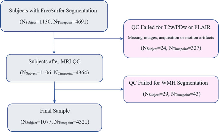
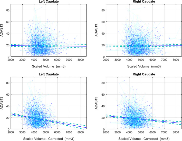
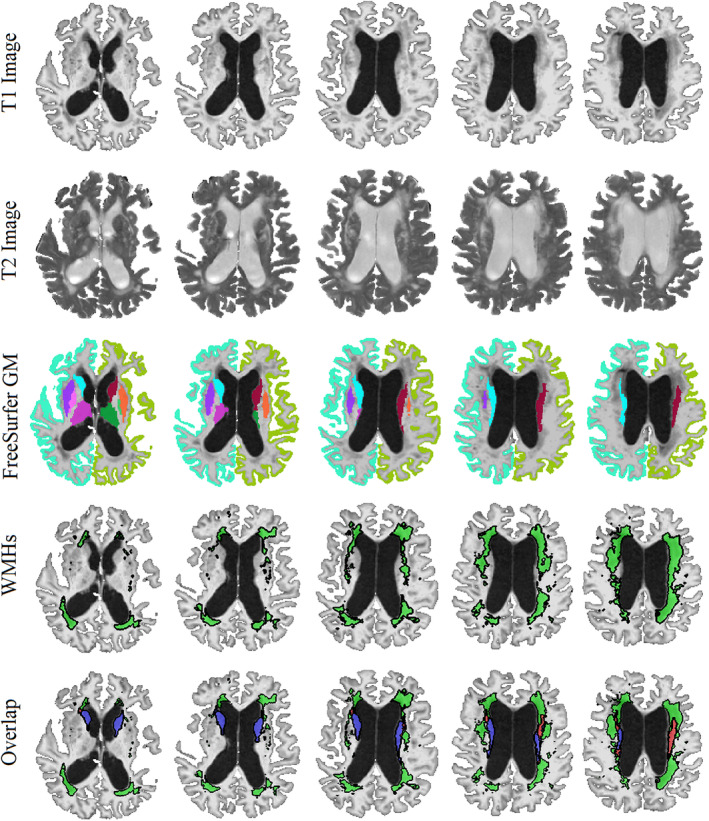
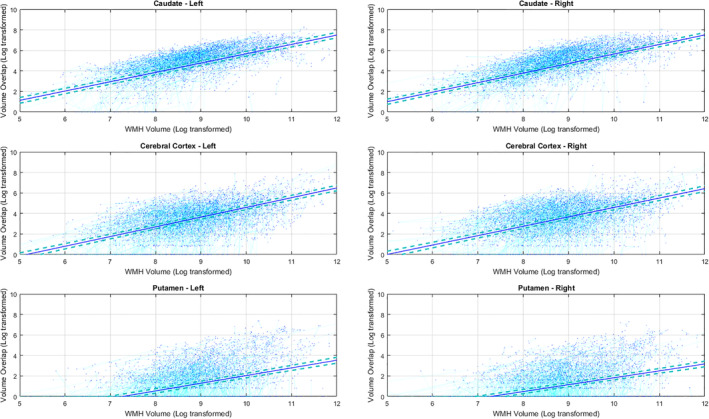
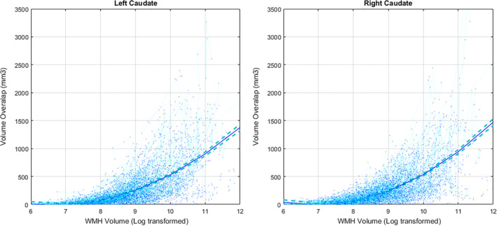
Volumetric estimates of subcortical and cortical structures, extracted from T1-weighted MRIs, are widely used in many clinical and research applications. Here, we investigate the impact of the presence of white matter hyperintensities (WMHs) on FreeSurfer gray matter (GM) structure volumes and its possible bias on functional relationships. T1-weighted images from 1,077 participants (4,321 timepoints) from the Alzheimer's Disease Neuroimaging Initiative were processed with FreeSurfer version 6.0.0. WMHs were segmented using a previously validated algorithm on either T2-weighted or Fluid-attenuated inversion recovery images. Mixed-effects models were used to assess the relationships between overlapping WMHs and GM structure volumes and overall WMH burden, as well as to investigate whether such overlaps impact associations with age, diagnosis, and cognitive performance. Participants with higher WMH volumes had higher overlaps with GM volumes of bilateral caudate, cerebral cortex, putamen, thalamus, pallidum, and accumbens areas (p < .0001). When not corrected for WMHs, caudate volumes increased with age (p < .0001) and were not different between cognitively healthy individuals and age-matched probable Alzheimer's disease patients. After correcting for WMHs, caudate volumes decreased with age (p < .0001), and Alzheimer's disease patients had lower caudate volumes than cognitively healthy individuals (p < .01). Uncorrected caudate volume was not associated with ADAS13 scores, whereas corrected lower caudate volumes were significantly associated with poorer cognitive performance (p < .0001). Presence of WMHs leads to systematic inaccuracies in GM segmentations, particularly for the caudate, which can also change clinical associations. While specifically measured for the Freesurfer toolkit, this problem likely affects other algorithms.
从 T1 加权磁共振成像中提取的皮质下和皮质结构的容积估计值在许多临床和研究应用中得到广泛应用。在这里,我们研究了脑白质高信号(WMHs)的存在对 Freesurfer 灰质(GM)结构体积的影响及其对功能关系的可能偏差。从阿尔茨海默病神经影像学倡议的 1077 名参与者(4321 个时间点)的 T1 加权图像中使用 Freesurfer 版本 6.0.0 进行处理。WMHs 是使用以前在 T2 加权或液体衰减反转恢复图像上验证的算法分割的。混合效应模型用于评估重叠 WMHs 与 GM 结构体积和总体 WMH 负担之间的关系,以及研究这些重叠是否会影响与年龄、诊断和认知表现的关联。WMH 体积较高的参与者与双侧尾状核、大脑皮层、壳核、丘脑、苍白球和伏隔核区域的 GM 体积重叠较高(p<0.0001)。当未校正 WMHs 时,尾状核体积随年龄增长而增加(p<0.0001),且在认知健康个体与年龄匹配的可能阿尔茨海默病患者之间无差异。校正 WMHs 后,尾状核体积随年龄下降(p<0.0001),且阿尔茨海默病患者的尾状核体积低于认知健康个体(p<0.01)。未校正的尾状核体积与 ADAS13 评分无关,而校正后的较低尾状核体积与较差的认知表现显著相关(p<0.0001)。WMHs 的存在导致 GM 分割的系统不准确,特别是对于尾状核,这也可能改变临床关联。虽然专门针对 Freesurfer 工具包进行了测量,但这个问题可能会影响其他算法。