• 文献检索
  • 文档翻译
  • 深度研究
  • 学术资讯
  • Suppr Zotero 插件Zotero 插件
  • 邀请有礼
  • 套餐&价格
  • 历史记录
应用&插件
Suppr Zotero 插件Zotero 插件浏览器插件Mac 客户端Windows 客户端微信小程序
定价
高级版会员购买积分包购买API积分包
服务
文献检索文档翻译深度研究API 文档MCP 服务
关于我们
关于 Suppr公司介绍联系我们用户协议隐私条款
关注我们

Suppr 超能文献

核心技术专利:CN118964589B侵权必究
粤ICP备2023148730 号-1Suppr @ 2026

文献检索

告别复杂PubMed语法,用中文像聊天一样搜索,搜遍4000万医学文献。AI智能推荐,让科研检索更轻松。

立即免费搜索

文件翻译

保留排版,准确专业,支持PDF/Word/PPT等文件格式,支持 12+语言互译。

免费翻译文档

深度研究

AI帮你快速写综述,25分钟生成高质量综述,智能提取关键信息,辅助科研写作。

立即免费体验

在严重急性 SARS-CoV-2 感染患者中,激活的 CD8 T 细胞中 CCR4 的上调表明增强了肺部归巢。

Upregulation of CCR4 in activated CD8 T cells indicates enhanced lung homing in patients with severe acute SARS-CoV-2 infection.

机构信息

Department of Internal Medicine 5, Hematology, and Oncology, University Hospital Erlangen and Friedrich-Alexander-University Erlangen-Nürnberg, Erlangen, Germany.

Institute of Virology, University Hospital Erlangen and Friedrich-Alexander-University Erlangen-Nürnberg, Erlangen, Germany.

出版信息

Eur J Immunol. 2021 Jun;51(6):1436-1448. doi: 10.1002/eji.202049135. Epub 2021 Apr 19.

DOI:10.1002/eji.202049135
PMID:33784417
原文链接:https://pmc.ncbi.nlm.nih.gov/articles/PMC8250120/
Abstract

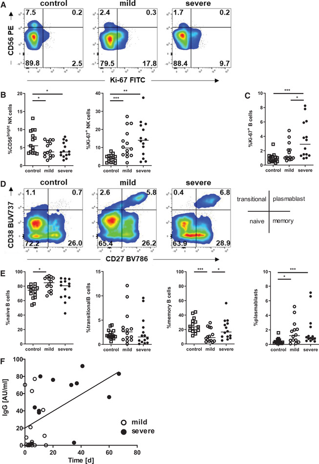
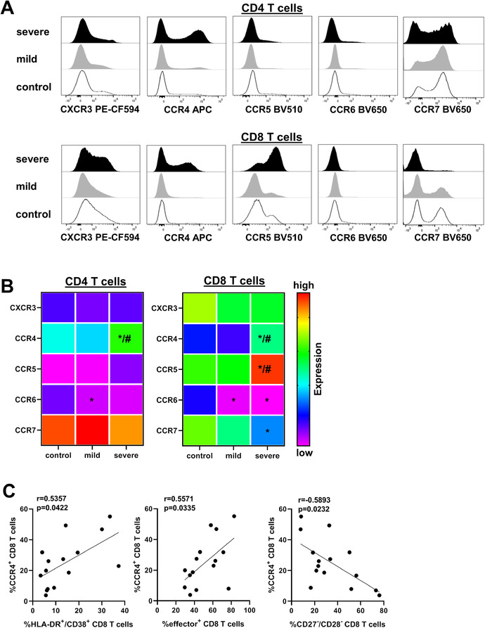
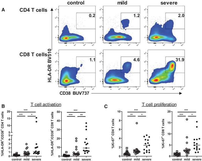
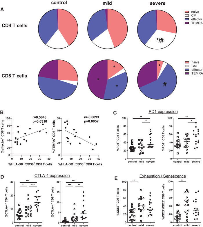
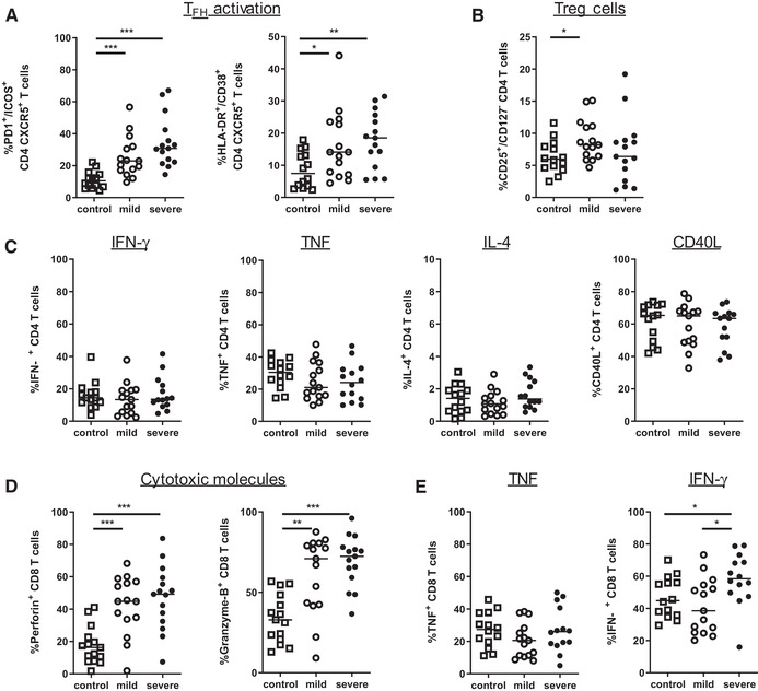
COVID-19 is a life-threatening disease leading to bilateral pneumonia and respiratory failure. The underlying reasons why a smaller percentage of patients present with severe pulmonary symptoms whereas the majority is only mildly affected are to date not well understood. Comparing the immunological phenotype in healthy donors and patients with mild versus severe COVID-19 shows that in COVID-19 patients, NK-/B-cell activation and proliferation are enhanced independent of severity. As an important precondition for effective antibody responses, T-follicular helper cells and antibody secreting cells are increased both in patients with mild and severe SARS-CoV-2 infection. Beyond this, T cells in COVID-19 patients exhibit a stronger activation profile with differentiation toward effector cell phenotypes. Importantly, when looking at the rates of pulmonary complications in COVID-19 patients, the chemokine receptor CCR4 is higher expressed by both CD4 and CD8 T cells of patients with severe COVID-19. This raises the hypothesis that CCR4 upregulation on T cells in the pathogenesis of COVID-19 promotes stronger T-cell attraction to the lungs leading to increased immune activation with presumably higher pulmonary toxicity. Our study contributes significantly to the understanding of the immunological changes during COVID-19, as new therapeutic agents, preferentially targeting the immune system, are highly warranted.

摘要

COVID-19 是一种危及生命的疾病,可导致双侧肺炎和呼吸衰竭。迄今为止,人们还不太清楚为什么只有一小部分患者出现严重的肺部症状,而大多数患者只是轻度感染的根本原因。比较健康供体与轻度和重度 COVID-19 患者的免疫表型表明,在 COVID-19 患者中,NK-/B 细胞的激活和增殖增强,而与严重程度无关。作为有效抗体反应的重要前提条件,滤泡辅助 T 细胞和分泌抗体的细胞在轻度和重度 SARS-CoV-2 感染的患者中均增加。除此之外,COVID-19 患者的 T 细胞表现出更强的激活特征,向效应细胞表型分化。重要的是,当观察 COVID-19 患者的肺部并发症发生率时,严重 COVID-19 患者的 CD4 和 CD8 T 细胞均高表达趋化因子受体 CCR4。这提出了一个假设,即在 COVID-19 的发病机制中,T 细胞上 CCR4 的上调促进了 T 细胞向肺部的更强吸引,从而导致免疫激活增加,可能导致更高的肺部毒性。我们的研究对理解 COVID-19 期间的免疫变化有重要贡献,因为需要开发新的治疗药物,特别是针对免疫系统的药物。

https://cdn.ncbi.nlm.nih.gov/pmc/blobs/c58c/8250120/adee8e852143/EJI-51--g001.jpg
https://cdn.ncbi.nlm.nih.gov/pmc/blobs/c58c/8250120/3b25b1dafd28/EJI-51--g002.jpg
https://cdn.ncbi.nlm.nih.gov/pmc/blobs/c58c/8250120/d1188ac70079/EJI-51--g004.jpg
https://cdn.ncbi.nlm.nih.gov/pmc/blobs/c58c/8250120/50daeae7e8cf/EJI-51--g006.jpg
https://cdn.ncbi.nlm.nih.gov/pmc/blobs/c58c/8250120/d2fd44ba6a27/EJI-51--g003.jpg
https://cdn.ncbi.nlm.nih.gov/pmc/blobs/c58c/8250120/adee8e852143/EJI-51--g001.jpg

相似文献

1
Upregulation of CCR4 in activated CD8 T cells indicates enhanced lung homing in patients with severe acute SARS-CoV-2 infection.在严重急性 SARS-CoV-2 感染患者中,激活的 CD8 T 细胞中 CCR4 的上调表明增强了肺部归巢。
Eur J Immunol. 2021 Jun;51(6):1436-1448. doi: 10.1002/eji.202049135. Epub 2021 Apr 19.
2
COVID-19 severity associates with pulmonary redistribution of CD1c+ DCs and inflammatory transitional and nonclassical monocytes.COVID-19 严重程度与 CD1c+ DCs 和炎症性过渡和非经典单核细胞在肺部的重新分布相关。
J Clin Invest. 2020 Dec 1;130(12):6290-6300. doi: 10.1172/JCI140335.
3
Negative Clinical Evolution in COVID-19 Patients Is Frequently Accompanied With an Increased Proportion of Undifferentiated Th Cells and a Strong Underrepresentation of the Th1 Subset.在 COVID-19 患者中,临床病情恶化常伴随着未分化 Th 细胞比例增加和 Th1 亚群明显减少。
Front Immunol. 2020 Nov 26;11:596553. doi: 10.3389/fimmu.2020.596553. eCollection 2020.
4
Dynamic SARS-CoV-2-Specific Immunity in Critically Ill Patients With Hypertension.急危重症高血压患者的 SARS-CoV-2 特异性动态免疫。
Front Immunol. 2020 Dec 10;11:596684. doi: 10.3389/fimmu.2020.596684. eCollection 2020.
5
PD-1-Expressing SARS-CoV-2-Specific CD8 T Cells Are Not Exhausted, but Functional in Patients with COVID-19.PD-1 表达的 SARS-CoV-2 特异性 CD8 T 细胞并未衰竭,而是在 COVID-19 患者中具有功能。
Immunity. 2021 Jan 12;54(1):44-52.e3. doi: 10.1016/j.immuni.2020.12.002. Epub 2020 Dec 14.
6
Lymphocyte Changes in Severe COVID-19: Delayed Over-Activation of STING?严重 COVID-19 中的淋巴细胞变化:STING 过度激活延迟?
Front Immunol. 2020 Dec 1;11:607069. doi: 10.3389/fimmu.2020.607069. eCollection 2020.
7
Pro-inflammatory microenvironment and systemic accumulation of CXCR3+ cell exacerbate lung pathology of old rhesus macaques infected with SARS-CoV-2.促炎微环境和 CXCR3+ 细胞的全身积累加剧了感染 SARS-CoV-2 的老年恒河猴的肺部病理。
Signal Transduct Target Ther. 2021 Sep 1;6(1):328. doi: 10.1038/s41392-021-00734-w.
8
Naturally activated adaptive immunity in COVID-19 patients.COVID-19 患者的天然激活适应性免疫。
J Cell Mol Med. 2020 Nov;24(21):12457-12463. doi: 10.1111/jcmm.15771. Epub 2020 Sep 25.
9
Unique immunological profile in patients with COVID-19.COVID-19 患者的独特免疫特征。
Cell Mol Immunol. 2021 Mar;18(3):604-612. doi: 10.1038/s41423-020-00557-9. Epub 2020 Oct 15.
10
Characterization of T lymphocytes in severe COVID-19 patients.严重 COVID-19 患者 T 淋巴细胞特征。
J Med Virol. 2021 Sep;93(9):5608-5613. doi: 10.1002/jmv.27037. Epub 2021 May 3.

引用本文的文献

1
Macrophages foster anti-tumor immunity by ZEB1-dependent cytotoxic T cell chemoattraction.巨噬细胞通过ZEB1依赖的细胞毒性T细胞趋化作用促进抗肿瘤免疫。
Commun Biol. 2025 Jul 1;8(1):976. doi: 10.1038/s42003-025-08339-7.
2
Mass Cytometry Analysis of High-Dimensional Single-Cell Immune Profiles in ZF2001-Vaccinated Patients Infected with SARS-CoV-2.对感染SARS-CoV-2的ZF2001疫苗接种患者的高维单细胞免疫图谱进行质谱流式细胞术分析。
Infect Drug Resist. 2025 May 14;18:2497-2509. doi: 10.2147/IDR.S511936. eCollection 2025.
3
Effect of delayed isolation of peripheral blood mononuclear cells on cell viability and functionality.

本文引用的文献

1
Rapid production of clinical-grade SARS-CoV-2 specific T cells.临床级严重急性呼吸综合征冠状病毒2特异性T细胞的快速生产。
Adv Cell Gene Ther. 2020 Oct;3(4):e101. doi: 10.1002/acg2.101. Epub 2020 Jul 31.
2
SARS-CoV-2-specific T cell responses and correlations with COVID-19 patient predisposition.SARS-CoV-2 特异性 T 细胞应答与 COVID-19 患者易感性的相关性。
J Clin Invest. 2020 Dec 1;130(12):6477-6489. doi: 10.1172/JCI140965.
3
Immune Alterations in a Patient with SARS-CoV-2-Related Acute Respiratory Distress Syndrome.COVID-19 相关急性呼吸窘迫综合征患者的免疫改变。
外周血单个核细胞延迟分离对细胞活力和功能的影响。
BMC Immunol. 2025 Mar 15;26(1):21. doi: 10.1186/s12865-025-00701-y.
4
NFAT single-deficient murine T cells reduce the risk of aGvHD while controlling cytomegalovirus infection.NFAT单缺陷小鼠T细胞在控制巨细胞病毒感染的同时降低了急性移植物抗宿主病的风险。
iScience. 2025 Feb 1;28(2):111937. doi: 10.1016/j.isci.2025.111937. eCollection 2025 Feb 21.
5
Construction and validation of a risk prediction model for postoperative lung infection in elderly patients with lung cancer.构建并验证老年肺癌患者术后肺部感染风险预测模型。
Medicine (Baltimore). 2024 Nov 1;103(44):e40337. doi: 10.1097/MD.0000000000040337.
6
Bronchial Asthma and COVID-19: Etiology, Pathological Triggers, and Therapeutic Considerations.支气管哮喘与2019冠状病毒病:病因、病理触发因素及治疗考量
Pathophysiology. 2024 May 27;31(2):269-287. doi: 10.3390/pathophysiology31020020.
7
Delineating immune variation between adult and children COVID-19 cases and associations with disease severity.阐明成人和儿童 COVID-19 病例之间的免疫差异及其与疾病严重程度的关联。
Sci Rep. 2024 Mar 1;14(1):5090. doi: 10.1038/s41598-024-55148-9.
8
Sarcoidosis-related autoimmune inflammation in COVID-19 convalescent patients.新冠康复患者中与结节病相关的自身免疫性炎症
Front Med (Lausanne). 2023 Dec 21;10:1271198. doi: 10.3389/fmed.2023.1271198. eCollection 2023.
9
The characterization of CD8 T-cell responses in COVID-19.新型冠状病毒肺炎患者 CD8 T 细胞应答特征。
Emerg Microbes Infect. 2024 Dec;13(1):2287118. doi: 10.1080/22221751.2023.2287118. Epub 2024 Jan 11.
10
Insights on Covid-19 with superimposed pulmonary histoplasmosis: The possible nexus.新冠合并肺组织胞浆菌病的研究进展:可能存在的关联。
Immun Inflamm Dis. 2023 Sep;11(9):e989. doi: 10.1002/iid3.989.
J Clin Immunol. 2020 Nov;40(8):1082-1092. doi: 10.1007/s10875-020-00839-x. Epub 2020 Aug 22.
4
Natural killer cell immunotypes related to COVID-19 disease severity.与 COVID-19 疾病严重程度相关的自然杀伤细胞免疫表型。
Sci Immunol. 2020 Aug 21;5(50). doi: 10.1126/sciimmunol.abd6832.
5
Selective and cross-reactive SARS-CoV-2 T cell epitopes in unexposed humans.未暴露于 SARS-CoV-2 人群中的选择性和交叉反应性 T 细胞表位。
Science. 2020 Oct 2;370(6512):89-94. doi: 10.1126/science.abd3871. Epub 2020 Aug 4.
6
SARS-CoV-2-reactive T cells in healthy donors and patients with COVID-19.SARS-CoV-2 反应性 T 细胞在健康供体和 COVID-19 患者中的研究。
Nature. 2020 Nov;587(7833):270-274. doi: 10.1038/s41586-020-2598-9. Epub 2020 Jul 29.
7
Deep immune profiling of COVID-19 patients reveals distinct immunotypes with therapeutic implications.深度免疫剖析 COVID-19 患者,揭示具有治疗意义的不同免疫类型。
Science. 2020 Sep 4;369(6508). doi: 10.1126/science.abc8511. Epub 2020 Jul 15.
8
SARS-CoV-2-specific T cell immunity in cases of COVID-19 and SARS, and uninfected controls.COVID-19 和 SARS 病例以及未感染对照者的 SARS-CoV-2 特异性 T 细胞免疫。
Nature. 2020 Aug;584(7821):457-462. doi: 10.1038/s41586-020-2550-z. Epub 2020 Jul 15.
9
Immunological and inflammatory profiles in mild and severe cases of COVID-19.COVID-19 轻症和重症患者的免疫和炎症特征。
Nat Commun. 2020 Jul 8;11(1):3410. doi: 10.1038/s41467-020-17240-2.
10
Marked T cell activation, senescence, exhaustion and skewing towards TH17 in patients with COVID-19 pneumonia.COVID-19 肺炎患者中标记的 T 细胞激活、衰老、衰竭和向 TH17 的倾斜。
Nat Commun. 2020 Jul 6;11(1):3434. doi: 10.1038/s41467-020-17292-4.