Magnetic Resonance Unit, Department of Radiology, National Institute of Cardiology, ul. Alpejska 42, 04-628, Warsaw, Poland.
Department of Cardiology and Interventional Angiology, National Institute of Cardiology, Warsaw, Poland.
Sci Rep. 2021 Mar 30;11(1):7146. doi: 10.1038/s41598-021-86532-4.
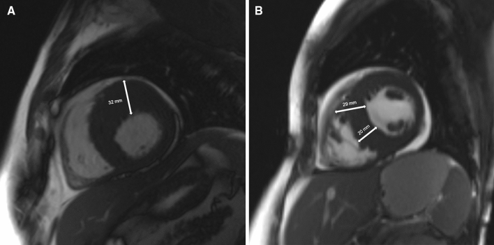
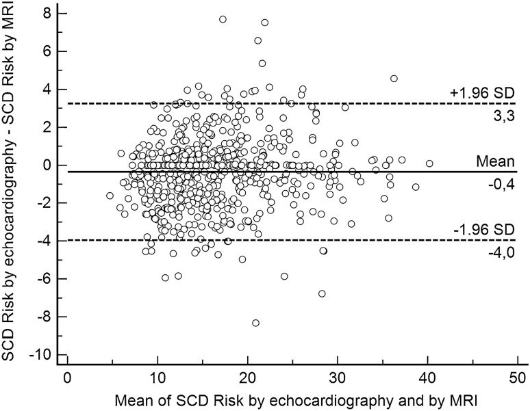
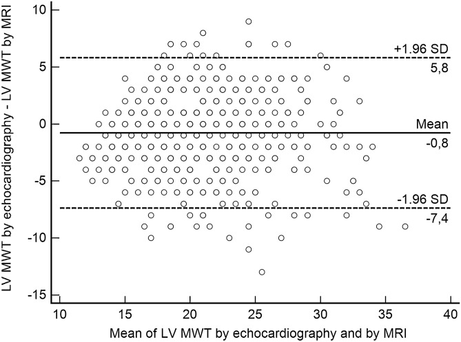
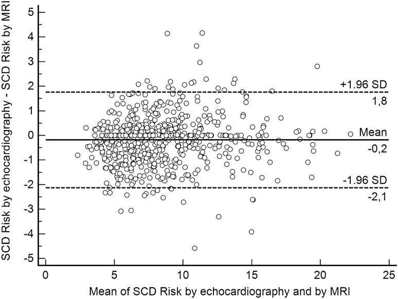
In hypertrophic cardiomyopathy (HCM) patients, left ventricular (LV) maximal wall thickness (MWT) is one of the most important factors determining sudden cardiac death (SCD) risk. In a large unselected sample of HCM patients, we aimed to simulate what changes would occur in the calculated SCD risk according to the European HCM Risk-SCD calculator when MWT measured using echocardiography was changed to MWT measured using MRI. All consecutive patients with HCM who underwent cardiac MRI were included. MWT measured with echocardiography and MRI were compared, and 5-year SCD risk according to the HCM Risk-SCD calculator was computed using four different models. The final population included 673 patients [389 (57.8%) males, median age 50 years, interquartile range (36-60)]. The median MWT was lower measured by echocardiography than by MRI [20 (17-24) mm vs 21 (18-24) mm; p < 0.0001]. There was agreement between echocardiography and MRI in the measurement of maximal LV wall thickness in 96 patients (14.3%). The largest differences between echo and MRI were - 13 mm and + 9 mm. The differences in MWT by echocardiography and MRI translated to a maximal difference of 8.33% in the absolute 5-year risk of SCD, i.e., the echocardiography-based risk was 8.33% lower than the MRI-based estimates. Interestingly, 13.7% of patients would have been reclassified into different SCD risk categories if MRI had been used to measure MWT instead of echocardiography. In conclusion, although there was high general intermodality agreement between echocardiography and MRI in the MWT measurements, the differences in MWT translated to significant differences in the 5-year risk of SCD.
在肥厚型心肌病(HCM)患者中,左心室(LV)最大壁厚度(MWT)是决定心脏性猝死(SCD)风险的最重要因素之一。在一项对大量未经选择的 HCM 患者的研究中,我们旨在模拟当使用 MRI 测量 MWT 代替超声心动图测量 MWT 时,欧洲 HCM 风险-SCD 计算器计算的 SCD 风险会发生什么变化。所有接受心脏 MRI 的连续 HCM 患者均被纳入研究。比较了超声心动图和 MRI 测量的 MWT,并使用 4 种不同的模型计算了 HCM Risk-SCD 计算器预测的 5 年 SCD 风险。最终纳入 673 例患者[389 例(57.8%)男性,中位年龄 50 岁,四分位间距 36-60]。超声心动图测量的 MWT 低于 MRI [20(17-24)mm 比 21(18-24)mm;p<0.0001]。在 96 例患者(14.3%)中,超声心动图和 MRI 对最大 LV 壁厚度的测量结果具有一致性。超声心动图和 MRI 之间的最大差异为-13mm 和+9mm。超声心动图和 MRI 测量的 MWT 差异导致 5 年 SCD 绝对风险的最大差异为 8.33%,即基于超声心动图的风险比基于 MRI 的估计低 8.33%。有趣的是,如果使用 MRI 测量 MWT 而不是超声心动图,13.7%的患者会被重新分类为不同的 SCD 风险类别。总之,尽管超声心动图和 MRI 之间的 MWT 测量具有高度的总体模态间一致性,但 MWT 的差异导致 5 年 SCD 风险的显著差异。