Oksdath Mansilla Mariana, Salazar-Hernandez Camilo, Perrin Sally L, Scheer Kaitlin G, Cildir Gökhan, Toubia John, Sedivakova Kristyna, Tea Melinda N, Lenin Sakthi, Ponthier Elise, Yeo Erica C F, Tergaonkar Vinay, Poonnoose Santosh, Ormsby Rebecca J, Pitson Stuart M, Brown Michael P, Ebert Lisa M, Gomez Guillermo A
Centre for Cancer Biology, SA Pathology and University of South Australia, Adelaide, SA, 5000, Australia.
ACRF Cancer Genomics Facility, Centre for Cancer Biology, SA Pathology and University of South Australia, Frome Road, Adelaide, SA, 5000, Australia.
BMC Biomed Eng. 2021 Apr 1;3(1):6. doi: 10.1186/s42490-021-00049-5.
Organoids are a reliable model used in the study of human brain development and under pathological conditions. However, current methods for brain organoid culture generate tissues that range from 0.5 to 2 mm of size, which need to be constantly agitated to allow proper oxygenation. The culture conditions are, therefore, not suitable for whole-brain organoid live imaging, required to study developmental processes and disease progression within physiologically relevant time frames (i.e. days, weeks, months).
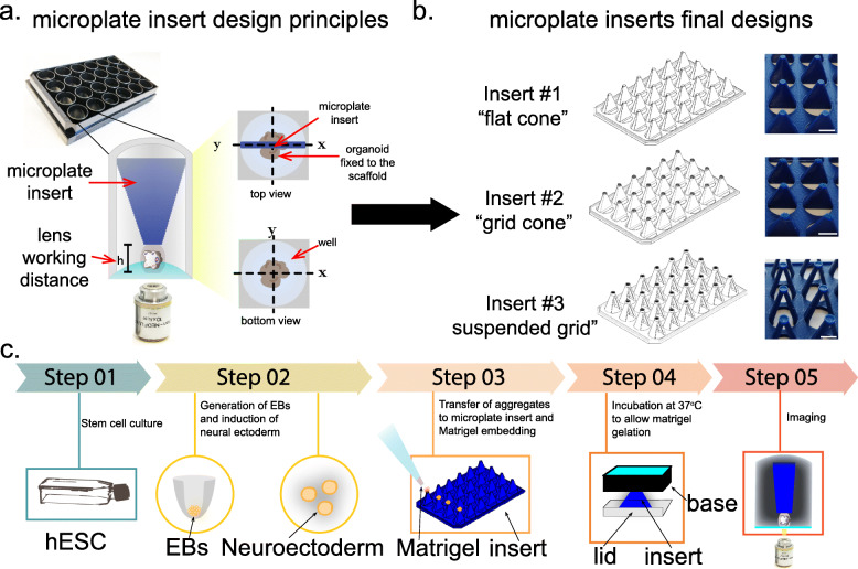
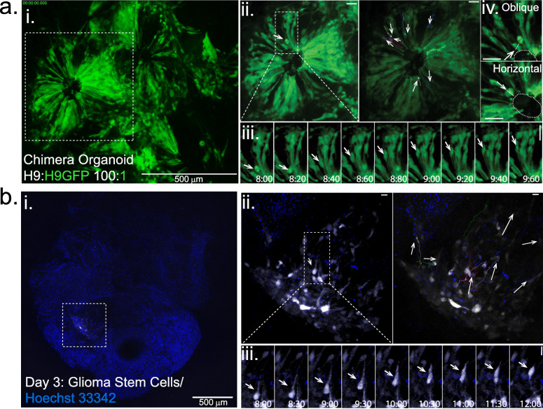
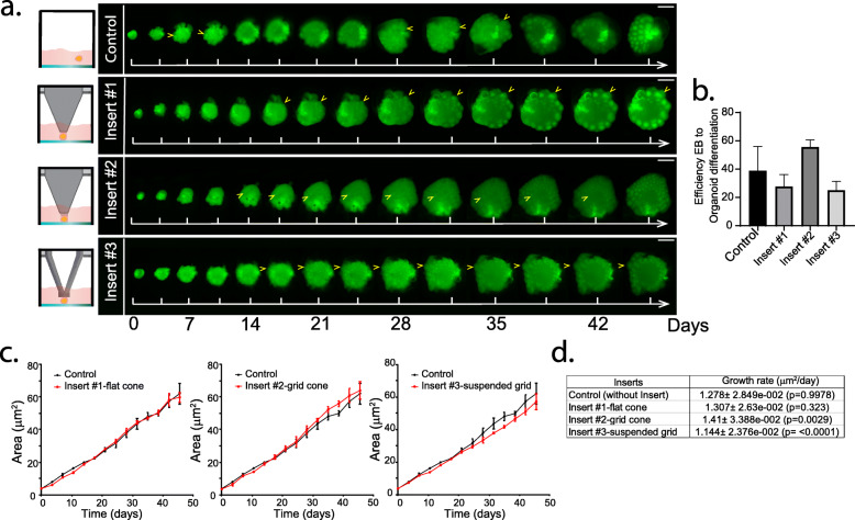
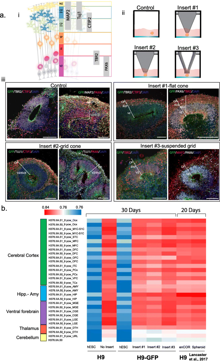
Here we designed 3D-printed microplate inserts adaptable to standard 24 multi-well plates, which allow the growth of multiple organoids in pre-defined and fixed XYZ coordinates. This innovation facilitates high-resolution imaging of whole-cerebral organoids, allowing precise assessment of organoid growth and morphology, as well as cell tracking within the organoids, over long periods. We applied this technology to track neocortex development through neuronal progenitors in brain organoids, as well as the movement of patient-derived glioblastoma stem cells within healthy brain organoids.
This new bioengineering platform constitutes a significant advance that permits long term detailed analysis of whole-brain organoids using multimodal inverted fluorescence microscopy.
类器官是用于研究人类大脑发育和病理状况的可靠模型。然而,目前的脑类器官培养方法所生成的组织大小在0.5到2毫米之间,需要不断搅拌以确保适当的氧合。因此,这种培养条件不适合全脑类器官的实时成像,而实时成像对于在生理相关的时间框架(即数天、数周、数月)内研究发育过程和疾病进展是必需的。
在此,我们设计了适用于标准24孔板的3D打印微孔板插入物,其允许多个类器官在预定义的固定XYZ坐标中生长。这一创新有助于对全脑类器官进行高分辨率成像,从而能够长期精确评估类器官的生长和形态,以及类器官内的细胞追踪。我们应用这项技术通过脑类器官中的神经祖细胞追踪新皮质发育,以及患者来源的胶质母细胞瘤干细胞在健康脑类器官内的运动。
这个新的生物工程平台是一项重大进展,它允许使用多模态倒置荧光显微镜对全脑类器官进行长期详细分析。