Rakotoson Irina, Delhomme Brigitte, Djian Philippe, Deeg Andreas, Brunstein Maia, Seebacher Christian, Uhl Rainer, Ricard Clément, Oheim Martin
Centre National de la Recherche Scientifique (CNRS) UMR 8118, Brain Physiology Laboratory, Paris, France.
Fédération de Recherche en Neurosciences CNRS FR 3636, Paris, France.
Front Neuroanat. 2019 Aug 20;13:77. doi: 10.3389/fnana.2019.00077. eCollection 2019.
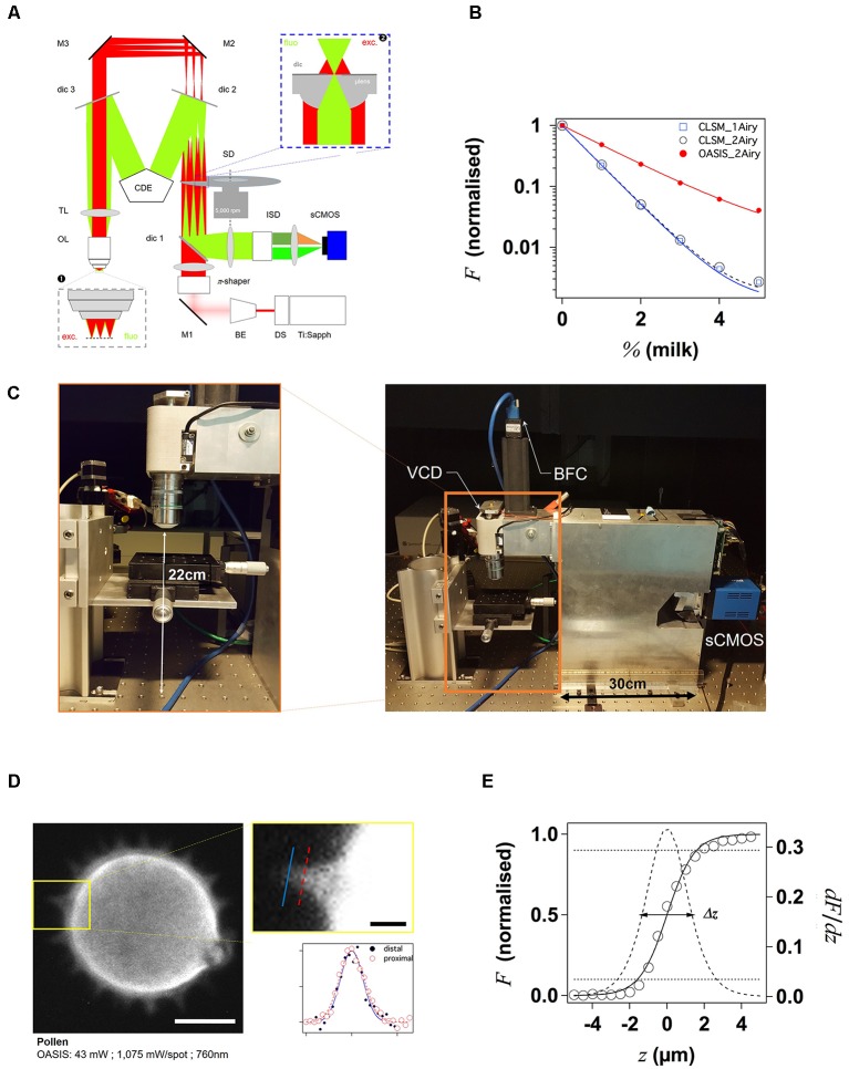
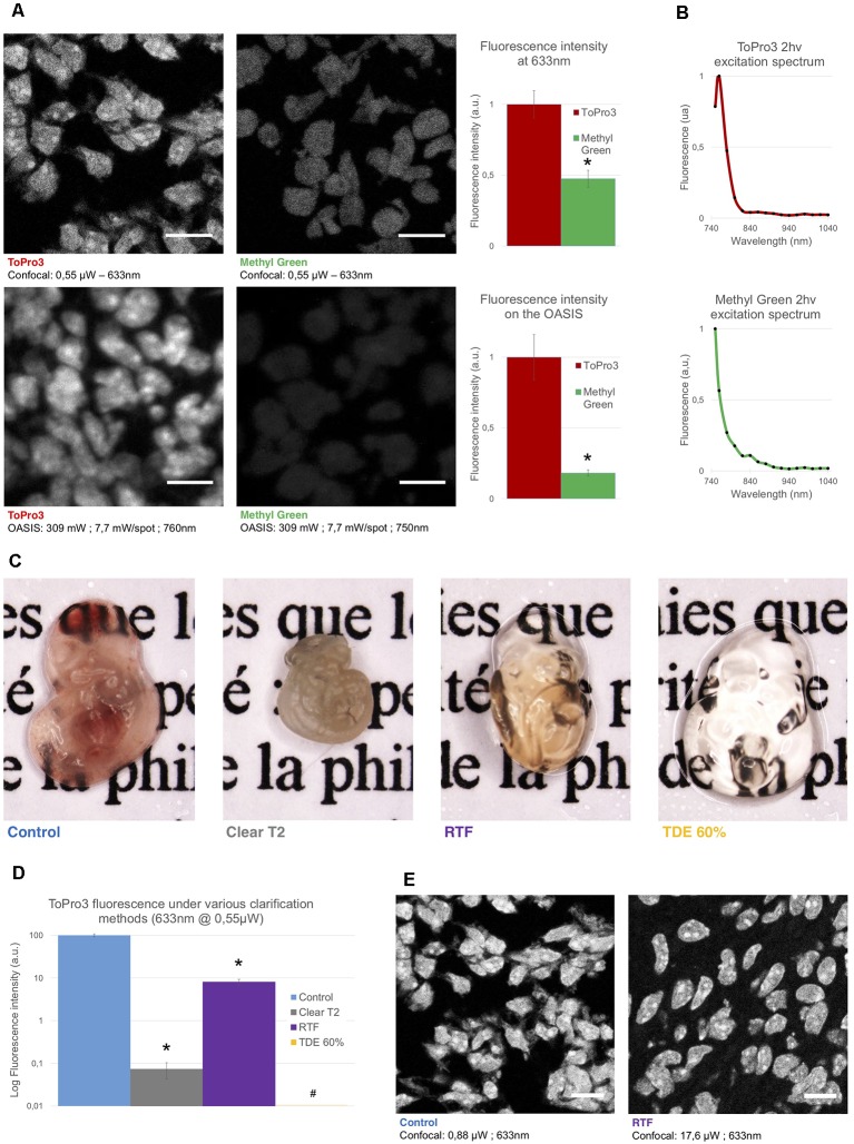
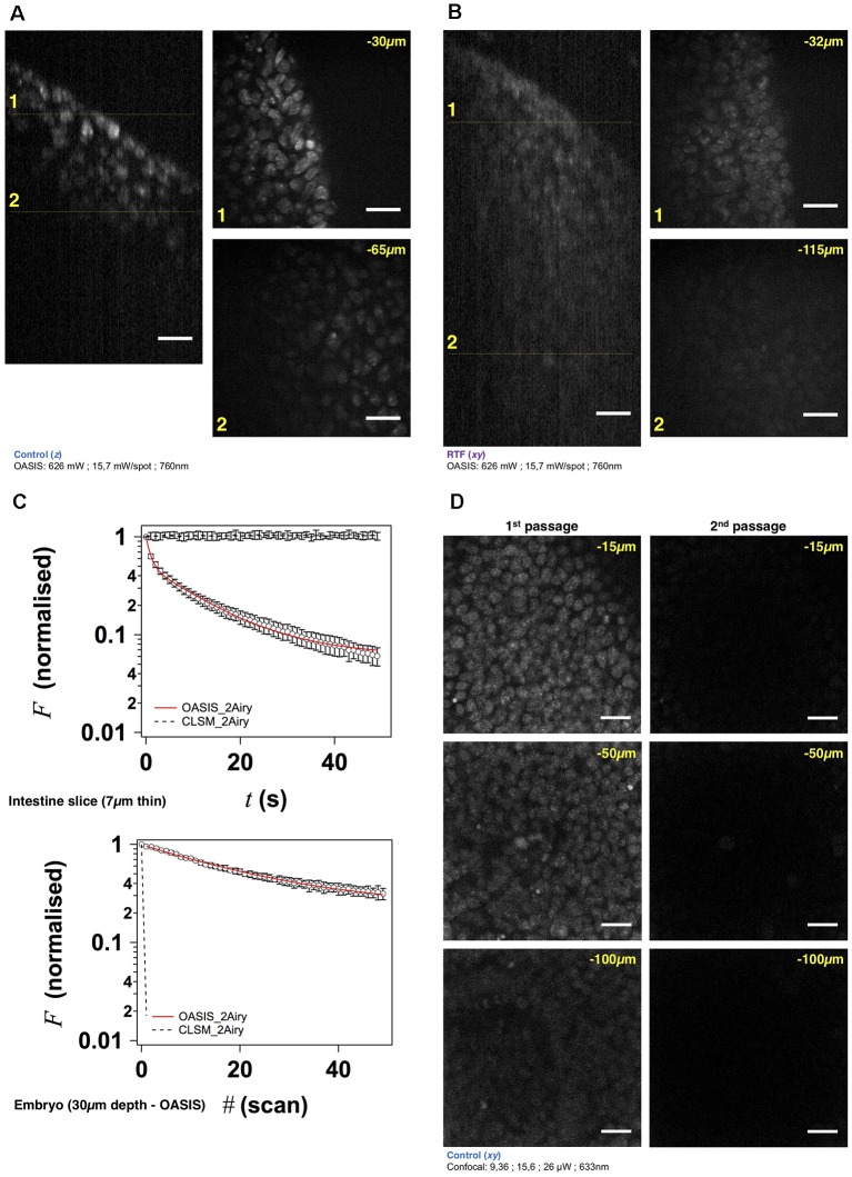
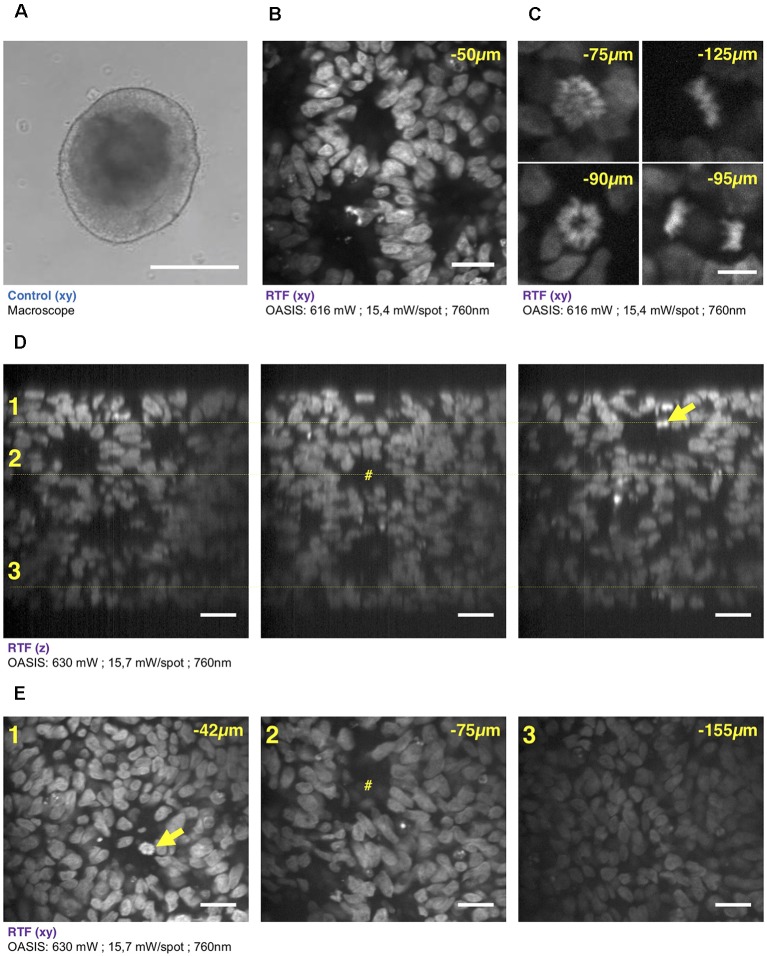
Human inducible pluripotent stem cells (hiPSCs) hold a large potential for disease modeling. hiPSC-derived human astrocyte and neuronal cultures permit investigations of neural signaling pathways with subcellular resolution. Combinatorial cultures, and three-dimensional (3-D) embryonic bodies (EBs) enlarge the scope of investigations to multi-cellular phenomena. The highest level of complexity, brain organoids that-in many aspects-recapitulate anatomical and functional features of the developing brain permit the study of developmental and morphological aspects of human disease. An ideal microscope for 3-D tissue imaging at these different scales would combine features from both confocal laser-scanning and light-sheet microscopes: a micrometric optical sectioning capacity and sub-micrometric spatial resolution, a large field of view and high frame rate, and a low degree of invasiveness, i.e., ideally, a better photon efficiency than that of a confocal microscope. In the present work, we describe such an instrument that uses planar two-photon (2P) excitation. Its particularity is that-unlike two- or three-lens light-sheet microscopes-it uses a single, low-magnification, high-numerical aperture objective for the generation and scanning of a virtual light sheet. The microscope builds on a modified Nipkow-Petráň spinning-disk scheme for achieving wide-field excitation. However, unlike the Yokogawa design that uses a tandem disk, our concept combines micro lenses, dichroic mirrors and detection pinholes on a single disk. This new design, advantageous for 2P excitation, circumvents problems arising with the tandem disk from the large wavelength difference between the infrared excitation light and visible fluorescence. 2P fluorescence excited by the light sheet is collected with the same objective and imaged onto a fast sCMOS camera. We demonstrate 3-D imaging of TO-PRO3-stained EBs and of brain organoids, uncleared and after rapid partial transparisation with triethanolamine formamide (RTF) and we compare the performance of our instrument to that of a confocal laser-scanning microscope (CLSM) having a similar numerical aperture. Our large-field 2P-spinning disk microscope permits one order of magnitude faster imaging, affords less photobleaching and permits better depth penetration than a confocal microscope with similar spatial resolution.
人类诱导多能干细胞(hiPSC)在疾病建模方面具有巨大潜力。源自hiPSC的人类星形胶质细胞和神经元培养物允许以亚细胞分辨率研究神经信号通路。组合培养和三维(3-D)胚胎体(EB)将研究范围扩大到多细胞现象。最高级别的复杂性,即脑类器官,在许多方面概括了发育中大脑的解剖和功能特征,允许研究人类疾病的发育和形态学方面。用于这些不同尺度的3-D组织成像的理想显微镜将结合共聚焦激光扫描显微镜和光片显微镜的特征:微米级光学切片能力和亚微米级空间分辨率、大视野和高帧率,以及低侵入性,即理想情况下,光子效率比共聚焦显微镜更高。在本工作中,我们描述了一种使用平面双光子(2P)激发的仪器。其独特之处在于,与双透镜或三透镜光片显微镜不同,它使用单个低倍率、高数值孔径物镜来生成和扫描虚拟光片。该显微镜基于改进的Nipkow-Petráň旋转盘方案实现宽场激发。然而,与使用串联盘的横河设计不同,我们的概念将微透镜、二向色镜和检测针孔组合在单个盘上。这种新设计有利于2P激发,避免了串联盘因红外激发光和可见荧光之间的大波长差异而产生的问题。由光片激发的2P荧光通过同一物镜收集并成像到快速sCMOS相机上。我们展示了用TO-PRO3染色的EB和脑类器官的3-D成像,未清除的以及用三乙醇胺甲酰胺(RTF)快速部分透明化后的成像,并将我们仪器的性能与具有相似数值孔径的共聚焦激光扫描显微镜(CLSM)的性能进行了比较。我们这种大视野2P旋转盘显微镜比具有相似空间分辨率的共聚焦显微镜成像速度快一个数量级,光漂白更少,深度穿透性更好。