Meira Willian, Daher Boutaina, Parks Scott Kenneth, Cormerais Yann, Durivault Jerome, Tambutte Eric, Pouyssegur Jacques, Vučetić Milica
Department of Medical Biology, Centre Scientifique de Monaco (CSM), 98000 Monaco, Monaco.
Trev and Joyce Deeley Research Centre, BC Cancer, Victoria, BC V8R 6V5, Canada.
Cancers (Basel). 2021 Mar 21;13(6):1434. doi: 10.3390/cancers13061434.
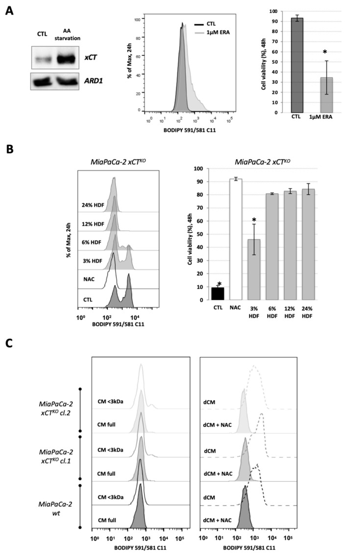
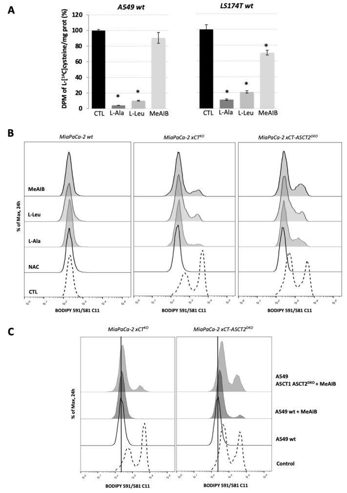
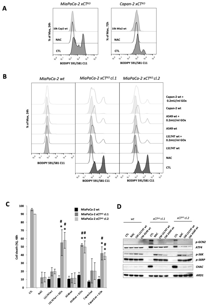
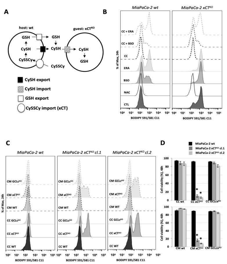
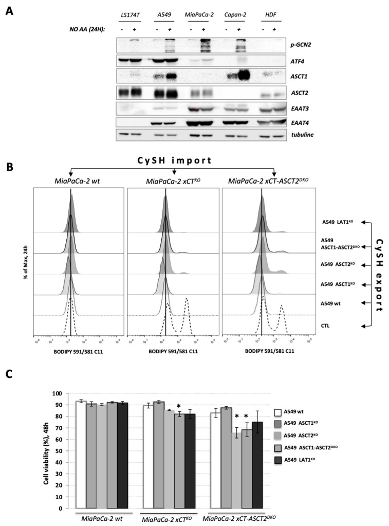
In our previous study, we showed that a cystine transporter (xCT) plays a pivotal role in ferroptosis of pancreatic ductal adenocarcinoma (PDAC) cells in vitro. However, in vivo xCT cells grew normally indicating that a mechanism exists to drastically suppress the ferroptotic phenotype. We hypothesized that plasma and neighboring cells within the tumor mass provide a source of cysteine to confer full ferroptosis resistance to xCT PDAC cells. To evaluate this hypothesis, we (co-) cultured xCT PDAC cells with different xCT-proficient cells or with their conditioned media. Our data unequivocally showed that the presence of a cysteine/cystine shuttle between neighboring cells is the mechanism that provides redox and nutrient balance, and thus ferroptotic resistance in xCT cells. Interestingly, although a glutathione shuttle between cells represents a good alternative hypothesis as a "rescue-mechanism", our data clearly demonstrated that the xCT phenotype is suppressed even with conditioned media from cells lacking the glutathione biosynthesis enzyme. Furthermore, we demonstrated that prevention of lipid hydroperoxide accumulation in vivo is mediated by import of cysteine into xCT cells via several genetically and pharmacologically identified transporters (ASCT1, ASCT2, LAT1, SNATs). Collectively, these data highlight the importance of the tumor environment in the ferroptosis sensitivity of cancer cells.
在我们之前的研究中,我们表明胱氨酸转运体(xCT)在体外胰腺导管腺癌(PDAC)细胞的铁死亡中起关键作用。然而,在体内xCT细胞生长正常,这表明存在一种机制可显著抑制铁死亡表型。我们推测血浆和肿瘤块内的邻近细胞提供了半胱氨酸来源,赋予xCT PDAC细胞完全的铁死亡抗性。为了评估这一假设,我们将xCT PDAC细胞与不同的xCT功能正常的细胞或其条件培养基(共)培养。我们的数据明确表明,邻近细胞之间存在半胱氨酸/胱氨酸穿梭机制,该机制提供氧化还原和营养平衡,从而赋予xCT细胞铁死亡抗性。有趣的是,尽管细胞间的谷胱甘肽穿梭作为一种“拯救机制”是一个很好的替代假设,但我们的数据清楚地表明,即使使用缺乏谷胱甘肽生物合成酶的细胞的条件培养基,xCT表型也会受到抑制。此外,我们证明了体内脂质氢过氧化物积累的预防是通过几种经基因和药理学鉴定的转运体(ASCT1、ASCT2、LAT1、SNATs)将半胱氨酸导入xCT细胞来介导的。总的来说,这些数据突出了肿瘤环境在癌细胞铁死亡敏感性中的重要性。