Department of Regional Pediatrics and Perinatology, Ehime University Graduate School of Medicine, Toon 791-0295, Japan.
Department of Obstetrics and Gynecology, Ehime University School of Medicine, Toon 791-0295, Japan.
Int J Mol Sci. 2021 Mar 4;22(5):2572. doi: 10.3390/ijms22052572.
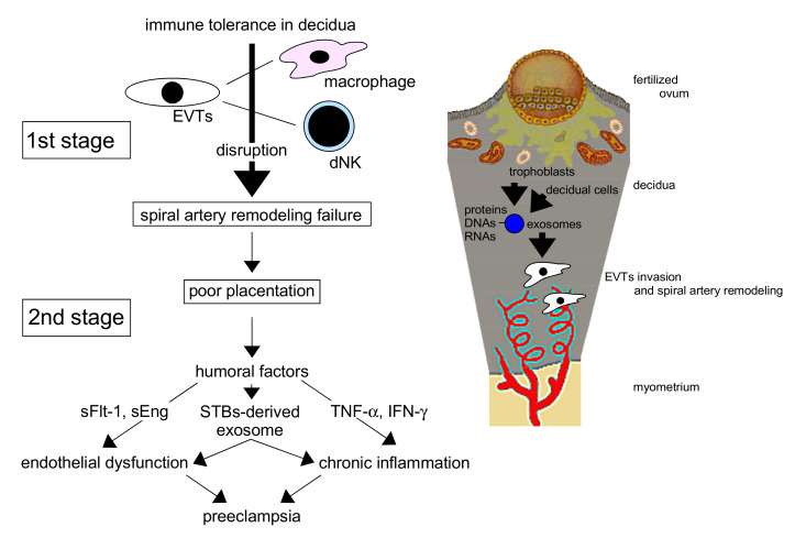
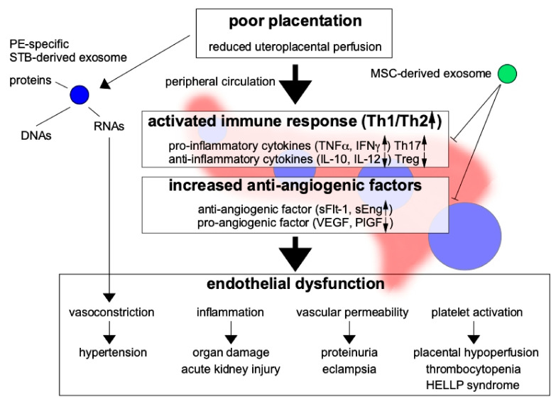
The pathogenesis of preeclampsia begins when a fertilized egg infiltrates the decidua, resulting in implantation failure (e.g., due to extravillous trophoblast infiltration disturbance and abnormal spiral artery remodeling). Thereafter, large amounts of serum factors (e.g., soluble fms-like tyrosine kinase 1 and soluble endoglin) are released into the blood from the hypoplastic placenta, and preeclampsia characterized by multiorgan disorder caused by vascular disorders develops. Successful implantation and placentation require immune tolerance to the fertilized egg as a semi-allograft and the stimulation of extravillous trophoblast infiltration. Recently, exosomes with diameters of 50-100 nm have been recognized to be involved in cell-cell communication. Exosomes affect cell functions in autocrine and paracrine manners via their encapsulating microRNA/DNA and membrane-bound proteins. The microRNA profiles of blood exosomes have been demonstrated to be useful for the evaluation of preeclampsia pathophysiology and prediction of the disease. In addition, exosomes derived from mesenchymal stem cells have been found to have cancer-suppressing effects. These exosomes may repair the pathophysiology of preeclampsia through the suppression of extravillous trophoblast apoptosis and promotion of these cells' invasive ability. Exosomes secreted by various cells have received much recent attention and may be involved in the maintenance of pregnancy and pathogenesis of preeclampsia.
子痫前期的发病机制始于受精卵浸润蜕膜,导致着床失败(例如,由于绒毛外滋养细胞浸润紊乱和异常螺旋动脉重塑)。此后,大量血清因子(例如,可溶性 fms 样酪氨酸激酶 1 和可溶性内皮糖蛋白)从发育不良的胎盘释放到血液中,由血管病变引起的多器官功能障碍的子痫前期随之发展。成功的着床和胎盘形成需要对受精卵作为半同种异体的免疫耐受以及绒毛外滋养细胞浸润的刺激。最近,直径为 50-100nm 的外泌体被认为参与细胞间通讯。外泌体通过其包裹的 microRNA/DNA 和膜结合蛋白以自分泌和旁分泌的方式影响细胞功能。血液外泌体的 microRNA 谱已被证明可用于评估子痫前期的病理生理学和预测疾病。此外,已发现间充质干细胞衍生的外泌体具有抑制癌症的作用。这些外泌体可能通过抑制绒毛外滋养细胞凋亡和促进这些细胞的侵袭能力来修复子痫前期的病理生理学。各种细胞分泌的外泌体最近受到了广泛关注,可能参与妊娠的维持和子痫前期的发病机制。