Kim Seung Hyeon, Kim Su-Jung, Park Jeongmin, Joe Yeonsoo, Lee So Eui, Saeidi Soma, Zhong Xiancai, Kim Seong Hoon, Park Sin-Aye, Na Hye-Kyung, Chung Hun Taeg, Surh Young-Joon
Cancer Research Institute, Seoul National University, Seoul 03087, Korea.
Research Institute of Pharmaceutical Sciences, College of Pharmacy, Seoul National University, Seoul 08826, Korea.
Antioxidants (Basel). 2021 Mar 16;10(3):470. doi: 10.3390/antiox10030470.
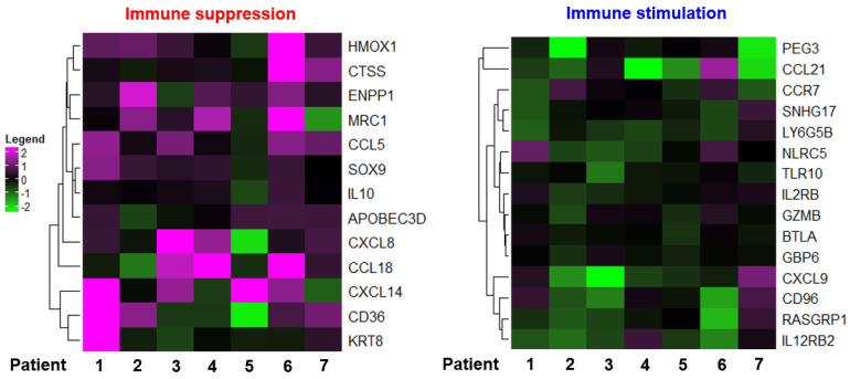
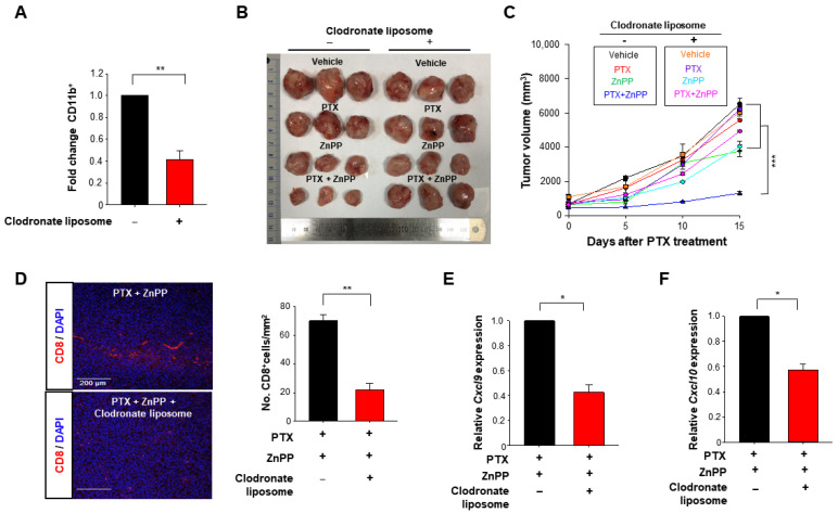
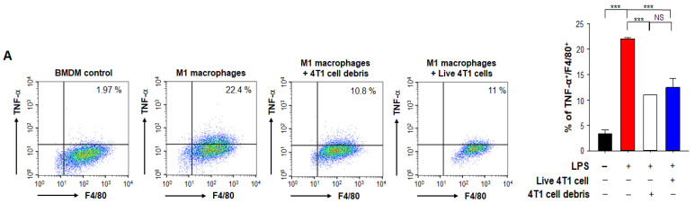
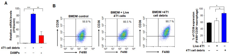
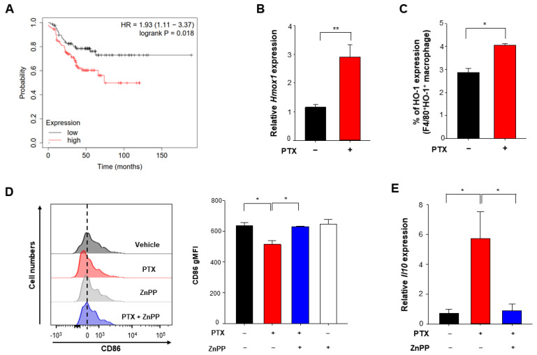
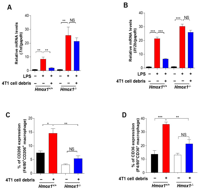
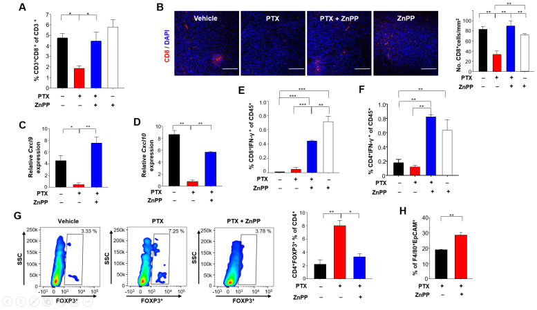
Tumor-associated macrophages (TAMs) represent one of the most abundant components of the tumor microenvironment and play important roles in tumor development and progression. TAMs display plasticity and functional heterogeneity as reflected by distinct phenotypic subsets. TAMs with an M1 phenotype have proinflammatory and anti-tumoral properties whereas M2-like TAMs exert anti-inflammatory and pro-tumoral functions. Tumor cell debris generated during chemotherapy can stimulate primary tumor growth and recurrence. According to our previous study, phagocytic engulfment of breast tumor cell debris by TAMs attenuated chemotherapeutic efficacy through the upregulation of heme oxygenase-1 (HO-1). To verify the impact of HO-1 upregulation on the profile of macrophage polarization during cytotoxic therapy, we utilized a syngeneic murine breast cancer (4T1) model in which tumor bearing mice were treated with paclitaxel (PTX). PTX treatment markedly downregulated the surface expression of the M1 marker CD86 in infiltrated TAMs. Notably, there were significantly more cytotoxic CD8 T cells in tumors of mice treated with PTX plus the HO-1 inhibitor, zinc protophorphyrin IX (ZnPP) than in mice treated with PTX alone. Interestingly, the tumor-inhibiting efficacy of PTX and ZnPP co-treatment was abrogated when macrophages were depleted by clodronate liposomes. Macrophage depletion also decreased the intratumoral CD8 T cell population and downregulated the expression of and The expression of the M1 phenotype marker, CD86 was higher in mice injected with PTX plus ZnPP than that in mice treated with PTX alone. Conversely, the PTX-induced upregulation of the M2 marker gene, in CD11b myeloid cells from 4T1 tumor-bearing mice treated was dramatically reduced by the administration of the HO-1 inhibitor. Genetic ablation of HO-1 abolished the inhibitory effect of 4T1 tumor cell debris on expression of M1 marker genes, and , in LPS-stimulated BMDMs. HO-1-deficient BMDMs exposed to tumor cell debris also exhibited a diminished expression of the M2 macrophage marker, CD206. These findings, taken all together, provide strong evidence that HO-1 plays a pivotal role in the transition of tumor-inhibiting M1-like TAMs to tumor-promoting M2-like ones during chemotherapy.
肿瘤相关巨噬细胞(TAMs)是肿瘤微环境中最丰富的成分之一,在肿瘤发生发展过程中发挥着重要作用。TAMs表现出可塑性和功能异质性,这通过不同的表型亚群得以体现。具有M1表型的TAMs具有促炎和抗肿瘤特性,而M2样TAMs则发挥抗炎和促肿瘤功能。化疗过程中产生的肿瘤细胞碎片可刺激原发性肿瘤生长和复发。根据我们之前的研究,TAMs对乳腺肿瘤细胞碎片的吞噬作用通过上调血红素加氧酶-1(HO-1)减弱了化疗疗效。为了验证HO-1上调对细胞毒性治疗期间巨噬细胞极化谱的影响,我们利用了同基因小鼠乳腺癌(4T1)模型,其中荷瘤小鼠接受紫杉醇(PTX)治疗。PTX治疗显著下调了浸润性TAMs中M1标志物CD86的表面表达。值得注意的是,与单独接受PTX治疗的小鼠相比,接受PTX加HO-1抑制剂锌原卟啉IX(ZnPP)治疗的小鼠肿瘤中具有细胞毒性的CD8 T细胞明显更多。有趣的是,当用氯膦酸脂质体清除巨噬细胞时,PTX和ZnPP联合治疗的肿瘤抑制效果被消除。巨噬细胞清除还减少了肿瘤内CD8 T细胞群体,并下调了 和 的表达。在注射PTX加ZnPP的小鼠中,M1表型标志物CD86的表达高于单独接受PTX治疗的小鼠。相反,给予HO-1抑制剂可显著降低PTX诱导的4T1荷瘤小鼠CD11b髓样细胞中M2标志物基因 的上调。HO-1基因敲除消除了4T1肿瘤细胞碎片对LPS刺激的骨髓来源巨噬细胞(BMDMs)中M1标志物基因 和 表达的抑制作用。暴露于肿瘤细胞碎片的HO-1缺陷型BMDMs也表现出M2巨噬细胞标志物CD206的表达降低。综上所述,这些发现提供了强有力的证据,表明HO-1在化疗期间肿瘤抑制性M1样TAMs向肿瘤促进性M2样TAMs的转变中起关键作用。