Sukhija Arsh, Mahajan Mangal, Joshi Priscilla C, Dsouza John, Seth Nagesh D N, Patil Karamchand H
Department of Radiodiagnosis and Imaging, Bharati Vidyapeeth Deemed to be University Medical College and Hospital, Pune, Maharashtra, India.
Department of Community Medicine, Bharati Vidyapeeth Deemed to be University Medical College and Hospital, Pune, Maharashtra, India.
Indian J Radiol Imaging. 2021 Jan;31(Suppl 1):S87-S93. doi: 10.4103/ijri.IJRI_777_20. Epub 2021 Jan 23.
As the burden of COVID-19 enhances, the need of a fast and reliable screening method is imperative. Chest radiographs plays a pivotal role in rapidly triaging the patients. Unfortunately, in low-resource settings, there is a scarcity of trained radiologists.
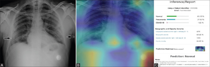
This study evaluates and compares the performance of an artificial intelligence (AI) system with a radiologist in detecting chest radiograph findings due to COVID-19.
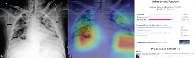
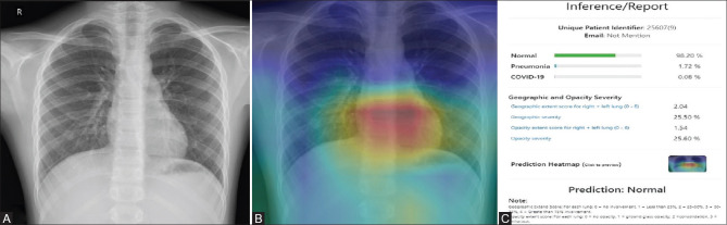
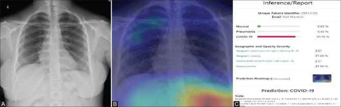
The test set consisted of 457 CXR images of patients with suspected COVID-19 pneumonia over a period of three months. The radiographs were evaluated by a radiologist with experience of more than 13 years and by the AI system (NeuraCovid, a web application that pairs with the AI model COVID-NET). Performance of AI system and the radiologist were compared by calculating the sensitivity, specificity and generating a receiver operating characteristic curve. RT-PCR test results were used as the gold standard.
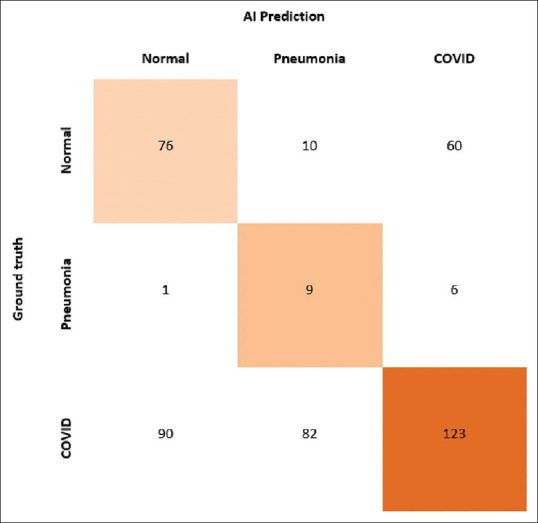
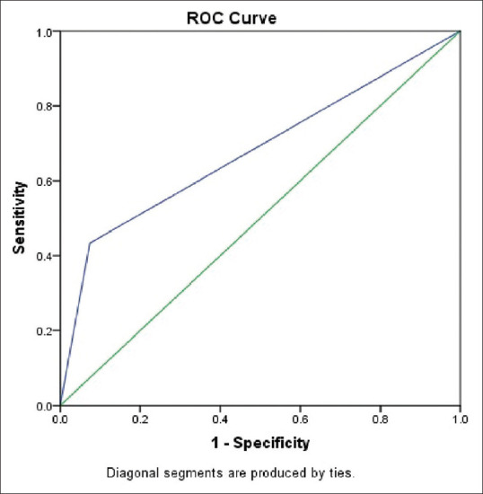
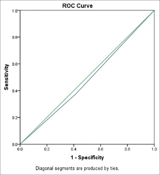
The radiologist obtained a sensitivity and specificity of 44.1% and 92.5%, respectively, whereas the AI had a sensitivity and specificity of 41.6% and 60%, respectively. The area under curve for correctly classifying CXR images as COVID-19 pneumonia was 0.48 for the AI system and 0.68 for the radiologist. The radiologist's prediction was found to be superior to that of the AI with a VALUE of .
The specificity and sensitivity of detecting lung involvement in COVID-19, by the radiologist, was found to be superior to that by the AI system.
随着新冠病毒病负担的加重,快速可靠的筛查方法势在必行。胸部X光片在对患者进行快速分诊中起着关键作用。不幸的是,在资源匮乏的地区,训练有素的放射科医生稀缺。
本研究评估并比较人工智能(AI)系统与放射科医生在检测新冠病毒病所致胸部X光片表现方面的性能。
测试集由三个月内457例疑似新冠病毒肺炎患者的胸部X光片图像组成。这些X光片由一位有超过13年经验的放射科医生和AI系统(NeuraCovid,一个与AI模型COVID-NET配对的网络应用程序)进行评估。通过计算灵敏度、特异度并生成受试者工作特征曲线来比较AI系统和放射科医生的性能。逆转录聚合酶链反应(RT-PCR)检测结果用作金标准。
放射科医生的灵敏度和特异度分别为44.1%和92.5%,而AI的灵敏度和特异度分别为41.6%和60%。将胸部X光片图像正确分类为新冠病毒肺炎的曲线下面积,AI系统为0.48,放射科医生为0.68。发现放射科医生的预测优于AI,其[具体数值]。
发现放射科医生在检测新冠病毒病肺部受累方面的特异度和灵敏度优于AI系统。