Dorr Francisco, Chaves Hernán, Serra María Mercedes, Ramirez Andrés, Costa Martín Elías, Seia Joaquín, Cejas Claudia, Castro Marcelo, Eyheremendy Eduardo, Fernández Slezak Diego, Farez Mauricio F
Entelai, Buenos Aires, Argentina.
Department of Diagnostic Imaging, Fleni, Buenos Aires, Argentina.
Intell Based Med. 2020 Dec;3:100014. doi: 10.1016/j.ibmed.2020.100014. Epub 2020 Nov 19.
To investigate the diagnostic performance of an Artificial Intelligence (AI) system for detection of COVID-19 in chest radiographs (CXR), and compare results to those of physicians working alone, or with AI support.
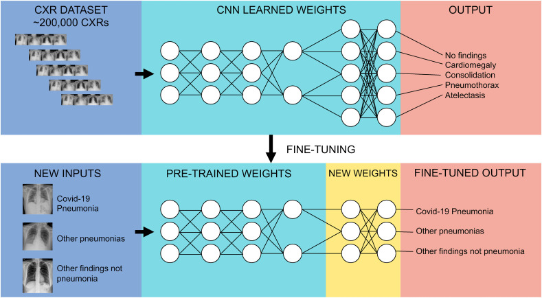
An AI system was fine-tuned to discriminate confirmed COVID-19 pneumonia, from other viral and bacterial pneumonia and non-pneumonia patients and used to review 302 CXR images from adult patients retrospectively sourced from nine different databases. Fifty-four physicians blind to diagnosis, were invited to interpret images under identical conditions in a test set, and randomly assigned either to receive or not receive support from the AI system. Comparisons were then made between diagnostic performance of physicians working with and without AI support. AI system performance was evaluated using the area under the receiver operating characteristic (AUROC), and sensitivity and specificity of physician performance compared to that of the AI system.
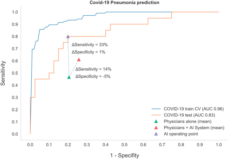
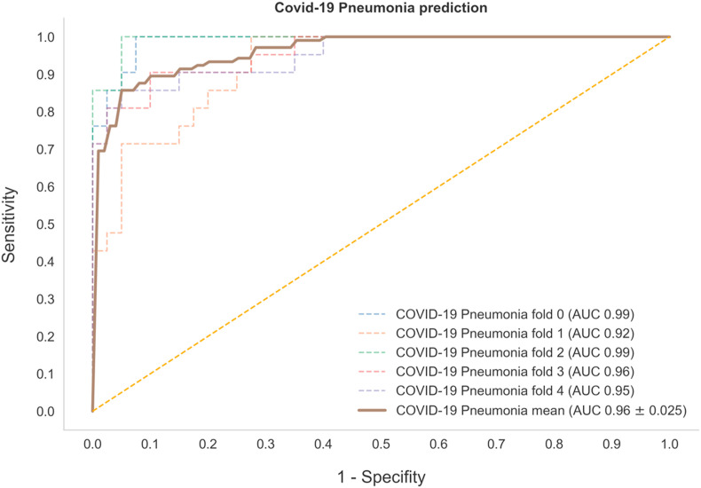
Discrimination by the AI system of COVID-19 pneumonia showed an AUROC curve of 0.96 in the validation and 0.83 in the external test set, respectively. The AI system outperformed physicians in the AUROC overall (70% increase in sensitivity and 1% increase in specificity, p < 0.0001). When working with AI support, physicians increased their diagnostic sensitivity from 47% to 61% (p < 0.001), although specificity decreased from 79% to 75% (p = 0.007).
Our results suggest interpreting chest radiographs (CXR) supported by AI, increases physician diagnostic sensitivity for COVID-19 detection. This approach involving a human-machine partnership may help expedite triaging efforts and improve resource allocation in the current crisis.
研究人工智能(AI)系统在胸部X光片(CXR)中检测新型冠状病毒肺炎(COVID-19)的诊断性能,并将结果与单独工作或有AI支持的医生的结果进行比较。
对一个AI系统进行微调,以区分确诊的COVID-19肺炎与其他病毒和细菌性肺炎以及非肺炎患者,并用于回顾性分析来自九个不同数据库的302例成年患者的CXR图像。邀请54名对诊断不知情的医生在相同条件下解读测试集中的图像,并随机分配是否接受AI系统的支持。然后比较有AI支持和无AI支持的医生的诊断性能。使用受试者操作特征曲线下面积(AUROC)评估AI系统的性能,并将医生的性能敏感性和特异性与AI系统进行比较。
AI系统对COVID-19肺炎的鉴别在验证集中的AUROC曲线为0.96,在外部测试集中为0.83。AI系统在AUROC方面总体上优于医生(敏感性提高70%,特异性提高1%,p<0.0001)。在有AI支持时,医生的诊断敏感性从47%提高到61%(p<0.001),尽管特异性从79%降至75%(p=0.007)。
我们的结果表明,在AI支持下解读胸部X光片可提高医生对COVID-19检测的诊断敏感性。这种人机合作的方法可能有助于加快分流工作,并改善当前危机中的资源分配。