• 文献检索
  • 文档翻译
  • 深度研究
  • 学术资讯
  • Suppr Zotero 插件Zotero 插件
  • 邀请有礼
  • 套餐&价格
  • 历史记录
应用&插件
Suppr Zotero 插件Zotero 插件浏览器插件Mac 客户端Windows 客户端微信小程序
定价
高级版会员购买积分包购买API积分包
服务
文献检索文档翻译深度研究API 文档MCP 服务
关于我们
关于 Suppr公司介绍联系我们用户协议隐私条款
关注我们

Suppr 超能文献

核心技术专利:CN118964589B侵权必究
粤ICP备2023148730 号-1Suppr @ 2026

文献检索

告别复杂PubMed语法,用中文像聊天一样搜索,搜遍4000万医学文献。AI智能推荐,让科研检索更轻松。

立即免费搜索

文件翻译

保留排版,准确专业,支持PDF/Word/PPT等文件格式,支持 12+语言互译。

免费翻译文档

深度研究

AI帮你快速写综述,25分钟生成高质量综述,智能提取关键信息,辅助科研写作。

立即免费体验

多放射科医师人工智能引导 COVID-19 肺部疾病严重程度在胸部 X 光片上的分级的用户研究。

Multi-Radiologist User Study for Artificial Intelligence-Guided Grading of COVID-19 Lung Disease Severity on Chest Radiographs.

机构信息

Athinoula A. Martinos Center for Biomedical Imaging, Department of Radiology, Massachusetts General Hospital, Harvard Medical School, Boston, Massachusetts.

Division of Thoracic Imaging and Intervention, Department of Radiology, Massachusetts General Hospital, Harvard Medical School, Boston, Massachusetts.

出版信息

Acad Radiol. 2021 Apr;28(4):572-576. doi: 10.1016/j.acra.2021.01.016. Epub 2021 Jan 18.

DOI:10.1016/j.acra.2021.01.016
PMID:33485773
原文链接:https://pmc.ncbi.nlm.nih.gov/articles/PMC7813473/
Abstract

RATIONALE AND OBJECTIVES

Radiographic findings of COVID-19 pneumonia can be used for patient risk stratification; however, radiologist reporting of disease severity is inconsistent on chest radiographs (CXRs). We aimed to see if an artificial intelligence (AI) system could help improve radiologist interrater agreement.

MATERIALS AND METHODS

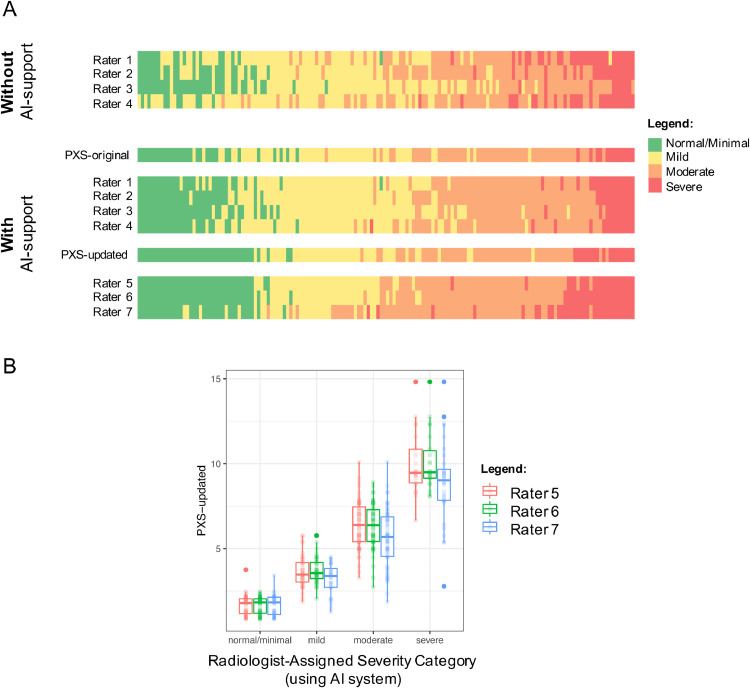
We performed a retrospective multi-radiologist user study to evaluate the impact of an AI system, the PXS score model, on the grading of categorical COVID-19 lung disease severity on 154 chest radiographs into four ordinal grades (normal/minimal, mild, moderate, and severe). Four radiologists (two thoracic and two emergency radiologists) independently interpreted 154 CXRs from 154 unique patients with COVID-19 hospitalized at a large academic center, before and after using the AI system (median washout time interval was 16 days). Three different thoracic radiologists assessed the same 154 CXRs using an updated version of the AI system trained on more imaging data. Radiologist interrater agreement was evaluated using Cohen and Fleiss kappa where appropriate. The lung disease severity categories were associated with clinical outcomes using a previously published outcomes dataset using Fisher's exact test and Chi-square test for trend.

RESULTS

Use of the AI system improved radiologist interrater agreement (Fleiss κ = 0.40 to 0.66, before and after use of the system). The Fleiss κ for three radiologists using the updated AI system was 0.74. Severity categories were significantly associated with subsequent intubation or death within 3 days.

CONCLUSION

An AI system used at the time of CXR study interpretation can improve the interrater agreement of radiologists.

摘要

背景和目的

COVID-19 肺炎的放射学表现可用于患者风险分层;然而,放射科医生对疾病严重程度的报告在胸部 X 线摄影(CXR)上不一致。我们旨在研究人工智能(AI)系统是否有助于提高放射科医生的组内一致性。

材料和方法

我们进行了一项回顾性多放射科医生用户研究,以评估 AI 系统(PXS 评分模型)对将 154 例 COVID-19 住院患者的 154 张胸部 X 线片的分类 COVID-19 肺部疾病严重程度分为四个有序等级(正常/最小、轻度、中度和重度)的影响。四位放射科医生(两位胸部放射科医生和两位急诊放射科医生)在使用 AI 系统前后(中位洗脱时间间隔为 16 天),分别独立解读了来自一家大型学术中心的 154 例 COVID-19 患者的 154 张 CXR。三位不同的胸部放射科医生使用经过更多影像学数据训练的更新版 AI 系统评估了相同的 154 张 CXR。使用 Cohen 和 Fleiss kappa 评估放射科医生的组内一致性,在适当的情况下。使用以前发表的使用 Fisher 确切检验和趋势卡方检验的结果数据集,将肺部疾病严重程度与临床结果相关联。

结果

使用 AI 系统提高了放射科医生的组内一致性(Fleiss κ 值从使用系统前后的 0.40 到 0.66)。三位放射科医生使用更新的 AI 系统的 Fleiss κ 值为 0.74。严重程度类别与 3 天内随后插管或死亡有显著相关性。

结论

在 CXR 研究解读时使用 AI 系统可以提高放射科医生的组内一致性。

https://cdn.ncbi.nlm.nih.gov/pmc/blobs/fbad/7813473/f020b386e697/gr2_lrg.jpg
https://cdn.ncbi.nlm.nih.gov/pmc/blobs/fbad/7813473/d0917d9e4fb1/gr1_lrg.jpg
https://cdn.ncbi.nlm.nih.gov/pmc/blobs/fbad/7813473/f020b386e697/gr2_lrg.jpg
https://cdn.ncbi.nlm.nih.gov/pmc/blobs/fbad/7813473/d0917d9e4fb1/gr1_lrg.jpg
https://cdn.ncbi.nlm.nih.gov/pmc/blobs/fbad/7813473/f020b386e697/gr2_lrg.jpg

相似文献

1
Multi-Radiologist User Study for Artificial Intelligence-Guided Grading of COVID-19 Lung Disease Severity on Chest Radiographs.多放射科医师人工智能引导 COVID-19 肺部疾病严重程度在胸部 X 光片上的分级的用户研究。
Acad Radiol. 2021 Apr;28(4):572-576. doi: 10.1016/j.acra.2021.01.016. Epub 2021 Jan 18.
2
Initial chest radiographs and artificial intelligence (AI) predict clinical outcomes in COVID-19 patients: analysis of 697 Italian patients.初始胸部 X 光片和人工智能 (AI) 预测 COVID-19 患者的临床结局:对 697 例意大利患者的分析。
Eur Radiol. 2021 Mar;31(3):1770-1779. doi: 10.1007/s00330-020-07269-8. Epub 2020 Sep 18.
3
Retrospectively assessing evaluation and management of artificial-intelligence detected nodules on uninterpreted chest radiographs in the era of radiologists shortage.回顾性评估在放射科医生短缺时代对未经解释的胸部 X 光片中人工智能检测到的结节的评估和管理。
Eur J Radiol. 2024 Jan;170:111241. doi: 10.1016/j.ejrad.2023.111241. Epub 2023 Nov 28.
4
COVID-19 pneumonia chest radiographic severity score: variability assessment among experienced and in-training radiologists and creation of a multireader composite score database for artificial intelligence algorithm development.COVID-19 肺炎胸部放射学严重程度评分:经验丰富的放射科医生和受训放射科医生之间的变异性评估,以及为人工智能算法开发创建多读者综合评分数据库。
Br J Radiol. 2022 Jun 1;95(1134):20211028. doi: 10.1259/bjr.20211028. Epub 2022 May 5.
5
Artificial Intelligence-assisted chest X-ray assessment scheme for COVID-19.人工智能辅助 COVID-19 胸片评估方案。
Eur Radiol. 2021 Aug;31(8):6039-6048. doi: 10.1007/s00330-020-07628-5. Epub 2021 Jan 20.
6
DeepCOVID-XR: An Artificial Intelligence Algorithm to Detect COVID-19 on Chest Radiographs Trained and Tested on a Large U.S. Clinical Data Set.DeepCOVID-XR:一种人工智能算法,可在美国大型临床数据集上进行训练和测试,用于检测胸部 X 光片上的 COVID-19。
Radiology. 2021 Apr;299(1):E167-E176. doi: 10.1148/radiol.2020203511. Epub 2020 Nov 24.
7
Radiologist-Trained AI Model for Identifying Suboptimal Chest-Radiographs.放射科医生培训 AI 模型以识别不优的胸部 X 光片。
Acad Radiol. 2023 Dec;30(12):2921-2930. doi: 10.1016/j.acra.2023.03.006. Epub 2023 Apr 3.
8
Artificial Intelligence Applied to Chest X-ray: A Reliable Tool to Assess the Differential Diagnosis of Lung Pneumonia in the Emergency Department.人工智能应用于胸部X光检查:急诊科评估肺炎鉴别诊断的可靠工具。
Diseases. 2023 Nov 20;11(4):171. doi: 10.3390/diseases11040171.
9
Chest radiograph classification and severity of suspected COVID-19 by different radiologist groups and attending clinicians: multi-reader, multi-case study.不同放射科医生组和临床医生对疑似 COVID-19 的胸部 X 线分类和严重程度:多读者、多病例研究。
Eur Radiol. 2023 Mar;33(3):2096-2104. doi: 10.1007/s00330-022-09172-w. Epub 2022 Oct 25.
10
Artificial intelligence-assisted double reading of chest radiographs to detect clinically relevant missed findings: a two-centre evaluation.人工智能辅助的胸部 X 光片双读以检测临床相关的遗漏发现:一项两中心评估。
Eur Radiol. 2024 Sep;34(9):5876-5885. doi: 10.1007/s00330-024-10676-w. Epub 2024 Mar 11.

引用本文的文献

1
MIDRC mRALE Mastermind Grand Challenge: AI to predict COVID severity on chest radiographs.MIDRC mRALE策划大师挑战赛:利用人工智能通过胸部X光片预测新冠严重程度。
J Med Imaging (Bellingham). 2025 Mar;12(2):024505. doi: 10.1117/1.JMI.12.2.024505. Epub 2025 Apr 18.
2
Expert-centered Evaluation of Deep Learning Algorithms for Brain Tumor Segmentation.专家对深度学习算法在脑肿瘤分割中的评估。
Radiol Artif Intell. 2024 Jan;6(1):e220231. doi: 10.1148/ryai.220231.
3
Artificial Intelligence Applied to Chest X-ray: A Reliable Tool to Assess the Differential Diagnosis of Lung Pneumonia in the Emergency Department.

本文引用的文献

1
Multi-population generalizability of a deep learning-based chest radiograph severity score for COVID-19.基于深度学习的 COVID-19 胸片严重程度评分在多人群中的泛化能力。
Medicine (Baltimore). 2022 Jul 22;101(29):e29587. doi: 10.1097/MD.0000000000029587.
2
Automated Assessment and Tracking of COVID-19 Pulmonary Disease Severity on Chest Radiographs using Convolutional Siamese Neural Networks.使用卷积暹罗神经网络对胸部X光片上的COVID-19肺部疾病严重程度进行自动评估和跟踪。
Radiol Artif Intell. 2020 Jul 22;2(4):e200079. doi: 10.1148/ryai.2020200079. eCollection 2020 Jul.
3
COVID-19 in CXR: From Detection and Severity Scoring to Patient Disease Monitoring.
人工智能应用于胸部X光检查:急诊科评估肺炎鉴别诊断的可靠工具。
Diseases. 2023 Nov 20;11(4):171. doi: 10.3390/diseases11040171.
4
A Hybrid Decision Tree and Deep Learning Approach Combining Medical Imaging and Electronic Medical Records to Predict Intubation Among Hospitalized Patients With COVID-19: Algorithm Development and Validation.一种结合医学影像和电子病历的混合决策树与深度学习方法,用于预测COVID-19住院患者的插管情况:算法开发与验证
JMIR Form Res. 2023 Oct 26;7:e46905. doi: 10.2196/46905.
5
Hospital-wide survey of clinical experience with artificial intelligence applied to daily chest radiographs.全院范围内对人工智能在日常胸部 X 光片中应用的临床经验调查。
PLoS One. 2023 Mar 2;18(3):e0282123. doi: 10.1371/journal.pone.0282123. eCollection 2023.
6
The 2021 SIIM-FISABIO-RSNA Machine Learning COVID-19 Challenge: Annotation and Standard Exam Classification of COVID-19 Chest Radiographs.2021 SIIM-FISABIO-RSNA 机器学习 COVID-19 挑战赛:COVID-19 胸部 X 光片的标注和标准考试分类。
J Digit Imaging. 2023 Feb;36(1):365-372. doi: 10.1007/s10278-022-00706-8. Epub 2022 Sep 28.
7
COVID-19 pneumonia chest radiographic severity score: variability assessment among experienced and in-training radiologists and creation of a multireader composite score database for artificial intelligence algorithm development.COVID-19 肺炎胸部放射学严重程度评分:经验丰富的放射科医生和受训放射科医生之间的变异性评估,以及为人工智能算法开发创建多读者综合评分数据库。
Br J Radiol. 2022 Jun 1;95(1134):20211028. doi: 10.1259/bjr.20211028. Epub 2022 May 5.
8
Diagnostic Test Accuracy of Deep Learning Detection of COVID-19: A Systematic Review and Meta-Analysis.深度学习检测 COVID-19 的诊断准确性:系统评价和荟萃分析。
Acad Radiol. 2021 Nov;28(11):1507-1523. doi: 10.1016/j.acra.2021.08.008. Epub 2021 Sep 17.
9
The Applications of Artificial Intelligence in Chest Imaging of COVID-19 Patients: A Literature Review.人工智能在COVID-19患者胸部成像中的应用:文献综述
Diagnostics (Basel). 2021 Jul 22;11(8):1317. doi: 10.3390/diagnostics11081317.
胸部X线片中的新型冠状病毒肺炎:从检测、严重程度评分到患者疾病监测
IEEE J Biomed Health Inform. 2021 Jun;25(6):1892-1903. doi: 10.1109/JBHI.2021.3069169. Epub 2021 Jun 3.
4
Automated Detection and Quantification of COVID-19 Airspace Disease on Chest Radiographs: A Novel Approach Achieving Expert Radiologist-Level Performance Using a Deep Convolutional Neural Network Trained on Digital Reconstructed Radiographs From Computed Tomography-Derived Ground Truth.基于 CT 图像构建的数字重建射线影像和真实标注的深度卷积神经网络在胸部 X 光片上对 COVID-19 肺部疾病的自动检测和量化:一种达到专家放射科医生水平的新方法。
Invest Radiol. 2021 Aug 1;56(8):471-479. doi: 10.1097/RLI.0000000000000763.
5
Initial chest radiographs and artificial intelligence (AI) predict clinical outcomes in COVID-19 patients: analysis of 697 Italian patients.初始胸部 X 光片和人工智能 (AI) 预测 COVID-19 患者的临床结局:对 697 例意大利患者的分析。
Eur Radiol. 2021 Mar;31(3):1770-1779. doi: 10.1007/s00330-020-07269-8. Epub 2020 Sep 18.
6
Predicting COVID-19 Pneumonia Severity on Chest X-ray With Deep Learning.利用深度学习通过胸部X光预测新冠肺炎肺炎严重程度
Cureus. 2020 Jul 28;12(7):e9448. doi: 10.7759/cureus.9448.
7
Determination of disease severity in COVID-19 patients using deep learning in chest X-ray images.利用胸部 X 光图像中的深度学习技术确定 COVID-19 患者的疾病严重程度。
Diagn Interv Radiol. 2021 Jan;27(1):20-27. doi: 10.5152/dir.2020.20205.
8
Deep transfer learning artificial intelligence accurately stages COVID-19 lung disease severity on portable chest radiographs.深度迁移学习人工智能可在便携式胸部 X 光片上准确分期 COVID-19 肺部疾病的严重程度。
PLoS One. 2020 Jul 28;15(7):e0236621. doi: 10.1371/journal.pone.0236621. eCollection 2020.
9
Dexamethasone in Hospitalized Patients with Covid-19.地塞米松在 COVID-19 住院患者中的应用。
N Engl J Med. 2021 Feb 25;384(8):693-704. doi: 10.1056/NEJMoa2021436. Epub 2020 Jul 17.
10
Racial and Ethnic Disparities in Disease Severity on Admission Chest Radiographs among Patients Admitted with Confirmed Coronavirus Disease 2019: A Retrospective Cohort Study.种族和民族差异对确诊 2019 年冠状病毒病患者入院时胸部 X 线片疾病严重程度的影响:一项回顾性队列研究。
Radiology. 2020 Dec;297(3):E303-E312. doi: 10.1148/radiol.2020202602. Epub 2020 Jul 16.