• 文献检索
  • 文档翻译
  • 深度研究
  • 学术资讯
  • Suppr Zotero 插件Zotero 插件
  • 邀请有礼
  • 套餐&价格
  • 历史记录
应用&插件
Suppr Zotero 插件Zotero 插件浏览器插件Mac 客户端Windows 客户端微信小程序
定价
高级版会员购买积分包购买API积分包
服务
文献检索文档翻译深度研究API 文档MCP 服务
关于我们
关于 Suppr公司介绍联系我们用户协议隐私条款
关注我们

Suppr 超能文献

核心技术专利:CN118964589B侵权必究
粤ICP备2023148730 号-1Suppr @ 2026

文献检索

告别复杂PubMed语法,用中文像聊天一样搜索,搜遍4000万医学文献。AI智能推荐,让科研检索更轻松。

立即免费搜索

文件翻译

保留排版,准确专业,支持PDF/Word/PPT等文件格式,支持 12+语言互译。

免费翻译文档

深度研究

AI帮你快速写综述,25分钟生成高质量综述,智能提取关键信息,辅助科研写作。

立即免费体验

新冠病毒病(COVID-19):一种用于分类的新型深度学习计算机辅助模型。

COVID-19: a new deep learning computer-aided model for classification.

作者信息

Elzeki Omar M, Shams Mahmoud, Sarhan Shahenda, Abd Elfattah Mohamed, Hassanien Aboul Ella

机构信息

Faculty of Computers and Information, Mansoura University, Mansoura, Egypt.

Faculty of Artificial Intelligence, Kafrelsheikh University, Kafrelsheikh, Egypt.

出版信息

PeerJ Comput Sci. 2021 Feb 18;7:e358. doi: 10.7717/peerj-cs.358. eCollection 2021.

DOI:10.7717/peerj-cs.358
PMID:33817008
原文链接:https://pmc.ncbi.nlm.nih.gov/articles/PMC7959596/
Abstract

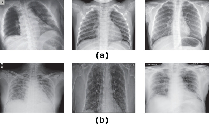
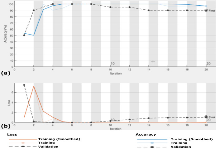
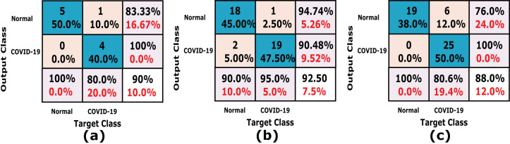
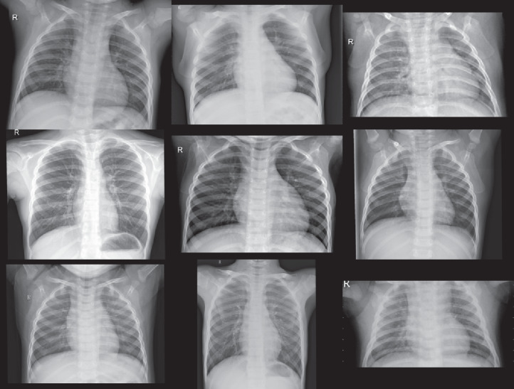
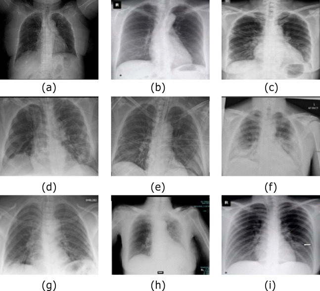
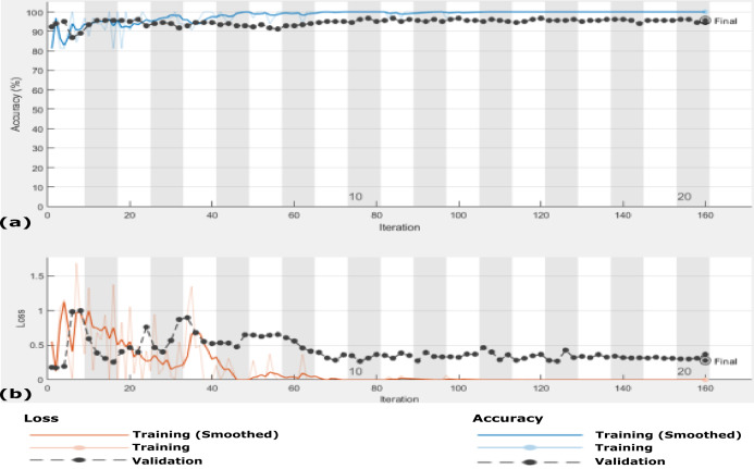
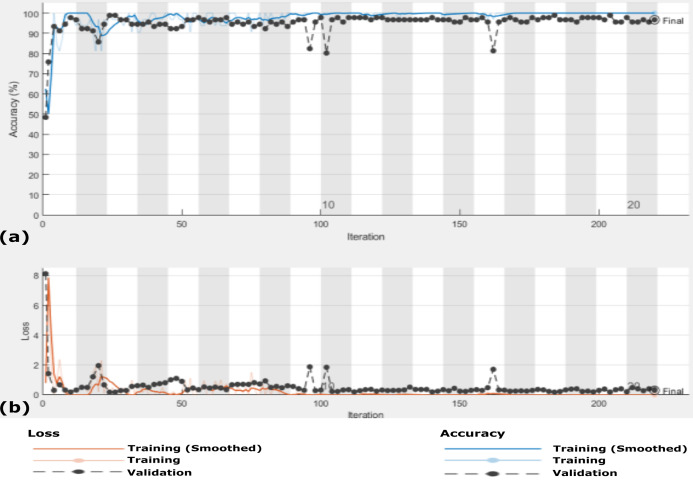
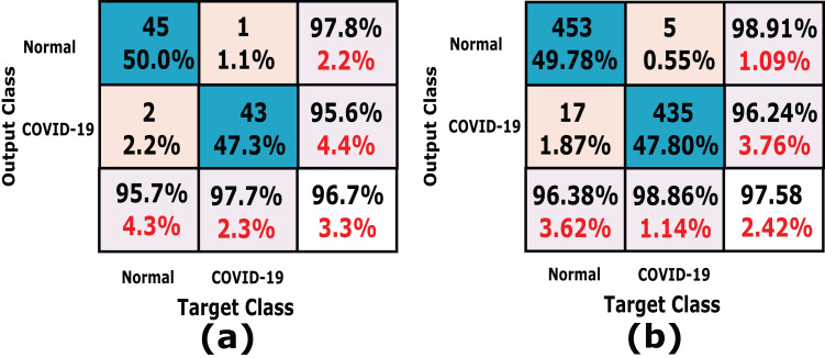
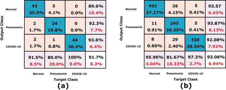
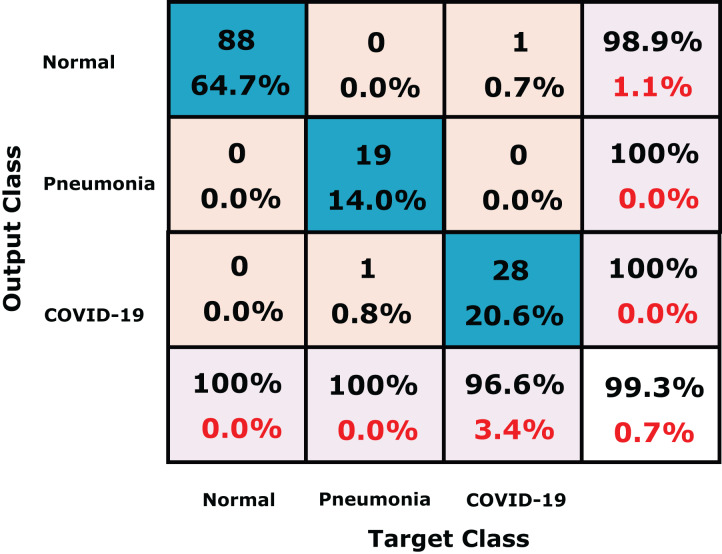
Chest X-ray (CXR) imaging is one of the most feasible diagnosis modalities for early detection of the infection of COVID-19 viruses, which is classified as a pandemic according to the World Health Organization (WHO) report in December 2019. COVID-19 is a rapid natural mutual virus that belongs to the coronavirus family. CXR scans are one of the vital tools to early detect COVID-19 to monitor further and control its virus spread. Classification of COVID-19 aims to detect whether a subject is infected or not. In this article, a model is proposed for analyzing and evaluating grayscale CXR images called Chest X-Ray COVID Network (CXRVN) based on three different COVID-19 X-Ray datasets. The proposed CXRVN model is a lightweight architecture that depends on a single fully connected layer representing the essential features and thus reducing the total memory usage and processing time verse pre-trained models and others. The CXRVN adopts two optimizers: mini-batch gradient descent and Adam optimizer, and the model has almost the same performance. Besides, CXRVN accepts CXR images in grayscale that are a perfect image representation for CXR and consume less memory storage and processing time. Hence, CXRVN can analyze the CXR image with high accuracy in a few milliseconds. The consequences of the learning process focus on decision making using a scoring function called SoftMax that leads to high rate true-positive classification. The CXRVN model is trained using three different datasets and compared to the pre-trained models: GoogleNet, ResNet and AlexNet, using the fine-tuning and transfer learning technologies for the evaluation process. To verify the effectiveness of the CXRVN model, it was evaluated in terms of the well-known performance measures such as precision, sensitivity, 1-score and accuracy. The evaluation results based on sensitivity, precision, recall, accuracy, and F1 score demonstrated that, after GAN augmentation, the accuracy reached 96.7% in experiment 2 (Dataset-2) for two classes and 93.07% in experiment-3 (Dataset-3) for three classes, while the average accuracy of the proposed CXRVN model is 94.5%.

摘要

胸部X光(CXR)成像是早期检测新型冠状病毒肺炎(COVID-19)病毒感染最可行的诊断方式之一,根据世界卫生组织(WHO)2019年12月的报告,COVID-19被列为大流行病。COVID-19是一种快速自然传播的病毒,属于冠状病毒家族。胸部X光扫描是早期检测COVID-19以进一步监测和控制其病毒传播的重要工具之一。COVID-19的分类旨在检测一个人是否被感染。在本文中,基于三个不同的COVID-19 X光数据集,提出了一种用于分析和评估灰度CXR图像的模型,称为胸部X光COVID网络(CXRVN)。所提出的CXRVN模型是一种轻量级架构,它依赖于一个表示基本特征的单一全连接层,从而减少了总内存使用量和处理时间,优于预训练模型和其他模型。CXRVN采用两种优化器:小批量梯度下降和Adam优化器,并且该模型具有几乎相同的性能。此外,CXRVN接受灰度的CXR图像,这是CXR的完美图像表示,占用更少的内存存储和处理时间。因此,CXRVN可以在几毫秒内高精度地分析CXR图像。学习过程的结果集中在使用称为SoftMax的评分函数进行决策,这导致了高比率的真阳性分类。CXRVN模型使用三个不同的数据集进行训练,并与预训练模型GoogleNet、ResNet和AlexNet进行比较,在评估过程中使用微调技术和迁移学习技术。为了验证CXRVN模型的有效性,根据精度、灵敏度、F1分数和准确率等著名的性能指标对其进行评估。基于灵敏度、精度、召回率、准确率和F1分数的评估结果表明,在进行生成对抗网络(GAN)增强后,在实验2(数据集2)中,两类的准确率达到96.7%,在实验3(数据集3)中三类的准确率达到93.07%,而所提出的CXRVN模型的平均准确率为94.5%。

https://cdn.ncbi.nlm.nih.gov/pmc/blobs/aad4/7959596/080a7ac408fc/peerj-cs-07-358-g015.jpg
https://cdn.ncbi.nlm.nih.gov/pmc/blobs/aad4/7959596/a2db89cb517f/peerj-cs-07-358-g001.jpg
https://cdn.ncbi.nlm.nih.gov/pmc/blobs/aad4/7959596/866e0e7510c2/peerj-cs-07-358-g002.jpg
https://cdn.ncbi.nlm.nih.gov/pmc/blobs/aad4/7959596/417512790485/peerj-cs-07-358-g003.jpg
https://cdn.ncbi.nlm.nih.gov/pmc/blobs/aad4/7959596/9875a71aa905/peerj-cs-07-358-g004.jpg
https://cdn.ncbi.nlm.nih.gov/pmc/blobs/aad4/7959596/f9d2f223c9b7/peerj-cs-07-358-g005.jpg
https://cdn.ncbi.nlm.nih.gov/pmc/blobs/aad4/7959596/ad0b96e21c90/peerj-cs-07-358-g006.jpg
https://cdn.ncbi.nlm.nih.gov/pmc/blobs/aad4/7959596/e0c43b5e36bd/peerj-cs-07-358-g007.jpg
https://cdn.ncbi.nlm.nih.gov/pmc/blobs/aad4/7959596/89f247c6179b/peerj-cs-07-358-g008.jpg
https://cdn.ncbi.nlm.nih.gov/pmc/blobs/aad4/7959596/e56eaf366c63/peerj-cs-07-358-g009.jpg
https://cdn.ncbi.nlm.nih.gov/pmc/blobs/aad4/7959596/1478df29ff98/peerj-cs-07-358-g010.jpg
https://cdn.ncbi.nlm.nih.gov/pmc/blobs/aad4/7959596/1b867ba91d34/peerj-cs-07-358-g011.jpg
https://cdn.ncbi.nlm.nih.gov/pmc/blobs/aad4/7959596/269b96dad86b/peerj-cs-07-358-g012.jpg
https://cdn.ncbi.nlm.nih.gov/pmc/blobs/aad4/7959596/c8d617f6754f/peerj-cs-07-358-g013.jpg
https://cdn.ncbi.nlm.nih.gov/pmc/blobs/aad4/7959596/c55956b793c0/peerj-cs-07-358-g014.jpg
https://cdn.ncbi.nlm.nih.gov/pmc/blobs/aad4/7959596/080a7ac408fc/peerj-cs-07-358-g015.jpg

相似文献

1
COVID-19: a new deep learning computer-aided model for classification.新冠病毒病(COVID-19):一种用于分类的新型深度学习计算机辅助模型。
PeerJ Comput Sci. 2021 Feb 18;7:e358. doi: 10.7717/peerj-cs.358. eCollection 2021.
2
Chest X-ray image phase features for improved diagnosis of COVID-19 using convolutional neural network.基于卷积神经网络的胸部 X 射线图像相位特征提高 COVID-19 诊断性能
Int J Comput Assist Radiol Surg. 2021 Feb;16(2):197-206. doi: 10.1007/s11548-020-02305-w. Epub 2021 Jan 9.
3
Development and integration of VGG and dense transfer-learning systems supported with diverse lung images for discovery of the Coronavirus identity.基于多种肺部图像支持的VGG和密集型迁移学习系统的开发与集成,用于冠状病毒特征发现。
Inform Med Unlocked. 2022;32:101004. doi: 10.1016/j.imu.2022.101004. Epub 2022 Jul 8.
4
COVID-19 detection in CT and CXR images using deep learning models.使用深度学习模型进行 CT 和 CXR 图像中的 COVID-19 检测。
Biogerontology. 2022 Feb;23(1):65-84. doi: 10.1007/s10522-021-09946-7. Epub 2022 Jan 22.
5
Automated image classification of chest X-rays of COVID-19 using deep transfer learning.利用深度迁移学习对新冠肺炎胸部X光片进行自动图像分类
Results Phys. 2021 Sep;28:104529. doi: 10.1016/j.rinp.2021.104529. Epub 2021 Jul 28.
6
Exploring the effect of image enhancement techniques on COVID-19 detection using chest X-ray images.探讨使用胸部 X 光图像的图像增强技术对 COVID-19 检测的影响。
Comput Biol Med. 2021 May;132:104319. doi: 10.1016/j.compbiomed.2021.104319. Epub 2021 Mar 11.
7
Computer-aided COVID-19 diagnosis and a comparison of deep learners using augmented CXRs.计算机辅助 COVID-19 诊断和使用增强 CXR 的深度学习方法比较。
J Xray Sci Technol. 2022;30(1):89-109. doi: 10.3233/XST-211047.
8
Pneumonia Classification Using Deep Learning from Chest X-ray Images During COVID-19.新冠疫情期间基于胸部X光图像利用深度学习进行肺炎分类
Cognit Comput. 2021 Jan 4:1-13. doi: 10.1007/s12559-020-09787-5.
9
Quantitative and Qualitative Analysis of 18 Deep Convolutional Neural Network (CNN) Models with Transfer Learning to Diagnose COVID-19 on Chest X-Ray (CXR) Images.基于迁移学习的18种深度卷积神经网络(CNN)模型对胸部X光(CXR)图像诊断新型冠状病毒肺炎(COVID-19)的定量和定性分析
SN Comput Sci. 2023;4(2):141. doi: 10.1007/s42979-022-01545-8. Epub 2023 Jan 5.
10
Deep Learning Algorithm for COVID-19 Classification Using Chest X-Ray Images.基于胸部 X 光图像的 COVID-19 分类深度学习算法。
Comput Math Methods Med. 2021 Nov 9;2021:9269173. doi: 10.1155/2021/9269173. eCollection 2021.

引用本文的文献

1
Challenges issues and future recommendations of deep learning techniques for SARS-CoV-2 detection utilising X-ray and CT images: a comprehensive review.利用X射线和CT图像进行SARS-CoV-2检测的深度学习技术面临的挑战、问题及未来建议:全面综述
PeerJ Comput Sci. 2024 Dec 24;10:e2517. doi: 10.7717/peerj-cs.2517. eCollection 2024.
2
Optimizing classification of diseases through language model analysis of symptoms.通过对症状进行语言模型分析来优化疾病分类。
Sci Rep. 2024 Jan 17;14(1):1507. doi: 10.1038/s41598-024-51615-5.
3
An augmentation aided concise CNN based architecture for COVID-19 diagnosis in real time.

本文引用的文献

1
Detection of COVID-19 using CXR and CT images using Transfer Learning and Haralick features.利用迁移学习和哈拉里克特征通过胸部X光和CT图像检测新型冠状病毒肺炎
Appl Intell (Dordr). 2021;51(1):341-358. doi: 10.1007/s10489-020-01831-z. Epub 2020 Aug 12.
2
Text Data Augmentation for Deep Learning.用于深度学习的文本数据增强
J Big Data. 2021;8(1):101. doi: 10.1186/s40537-021-00492-0. Epub 2021 Jul 19.
3
CovidGAN: Data Augmentation Using Auxiliary Classifier GAN for Improved Covid-19 Detection.CovidGAN:使用辅助分类器生成对抗网络进行数据增强以改进新冠病毒检测
一种基于增强辅助简洁 CNN 的实时 COVID-19 诊断架构。
Sci Rep. 2024 Jan 11;14(1):1136. doi: 10.1038/s41598-024-51317-y.
4
An Optimized Model Based on Deep Learning and Gated Recurrent Unit for COVID-19 Death Prediction.基于深度学习和门控循环单元的COVID-19死亡预测优化模型
Biomimetics (Basel). 2023 Nov 17;8(7):552. doi: 10.3390/biomimetics8070552.
5
COVID-SegNet: encoder-decoder-based architecture for COVID-19 lesion segmentation in chest X-ray.COVID-SegNet:用于胸部X光片中COVID-19病变分割的基于编码器-解码器的架构。
Multimed Syst. 2023 Apr 19:1-14. doi: 10.1007/s00530-023-01096-9.
6
COV-MobNets: a mobile networks ensemble model for diagnosis of COVID-19 based on chest X-ray images.COV-MobNets:一种基于胸部 X 光图像的用于 COVID-19 诊断的移动网络集成模型。
BMC Med Imaging. 2023 Jun 15;23(1):83. doi: 10.1186/s12880-023-01039-w.
7
Learning without forgetting by leveraging transfer learning for detecting COVID-19 infection from CT images.利用迁移学习从 CT 图像中检测 COVID-19 感染,实现无遗忘学习。
Sci Rep. 2023 May 25;13(1):8516. doi: 10.1038/s41598-023-34908-z.
8
BND-VGG-19: A deep learning algorithm for COVID-19 identification utilizing X-ray images.BND-VGG-19:一种利用X射线图像识别新冠肺炎的深度学习算法。
Knowl Based Syst. 2022 Dec 22;258:110040. doi: 10.1016/j.knosys.2022.110040. Epub 2022 Oct 21.
9
The effect of choosing optimizer algorithms to improve computer vision tasks: a comparative study.选择优化器算法对改进计算机视觉任务的影响:一项比较研究。
Multimed Tools Appl. 2023;82(11):16591-16633. doi: 10.1007/s11042-022-13820-0. Epub 2022 Sep 28.
10
COVID-19 Semantic Pneumonia Segmentation and Classification Using Artificial Intelligence.利用人工智能进行 COVID-19 语义肺炎分割和分类。
Contrast Media Mol Imaging. 2022 Sep 15;2022:5297709. doi: 10.1155/2022/5297709. eCollection 2022.
IEEE Access. 2020 May 14;8:91916-91923. doi: 10.1109/ACCESS.2020.2994762. eCollection 2020.
4
The importance of standardisation - COVID-19 CT & Radiograph Image Data Stock for deep learning purpose.标准化的重要性——用于深度学习目的的 COVID-19 CT 和 X 光图像数据资源库。
Comput Biol Med. 2020 Dec;127:104092. doi: 10.1016/j.compbiomed.2020.104092. Epub 2020 Oct 28.
5
Deep learning approaches for COVID-19 detection based on chest X-ray images.基于胸部X光图像的新冠肺炎检测深度学习方法
Expert Syst Appl. 2021 Feb;164:114054. doi: 10.1016/j.eswa.2020.114054. Epub 2020 Sep 28.
6
COVID-19 image classification using deep features and fractional-order marine predators algorithm.使用深度特征和分数阶海洋捕食者算法进行 COVID-19 图像分类。
Sci Rep. 2020 Sep 21;10(1):15364. doi: 10.1038/s41598-020-71294-2.
7
COVID-19 Prediction Models and Unexploited Data.COVID-19 预测模型和未开发数据。
J Med Syst. 2020 Aug 13;44(9):170. doi: 10.1007/s10916-020-01645-z.
8
Signs and symptoms to determine if a patient presenting in primary care or hospital outpatient settings has COVID-19 disease.用于确定在基层医疗或医院门诊就诊的患者是否患有新冠病毒病的体征和症状。
Cochrane Database Syst Rev. 2020 Jul 7;7(7):CD013665. doi: 10.1002/14651858.CD013665.
9
Truncated inception net: COVID-19 outbreak screening using chest X-rays.截断的 inception 网络:利用胸部 X 光进行 COVID-19 爆发筛查。
Phys Eng Sci Med. 2020 Sep;43(3):915-925. doi: 10.1007/s13246-020-00888-x. Epub 2020 Jun 25.
10
COVID-19 detection using deep learning models to exploit Social Mimic Optimization and structured chest X-ray images using fuzzy color and stacking approaches.利用深度学习模型进行 COVID-19 检测,利用模糊颜色和堆叠方法挖掘社会模拟优化和结构化胸部 X 射线图像。
Comput Biol Med. 2020 Jun;121:103805. doi: 10.1016/j.compbiomed.2020.103805. Epub 2020 May 6.