• 文献检索
  • 文档翻译
  • 深度研究
  • 学术资讯
  • Suppr Zotero 插件Zotero 插件
  • 邀请有礼
  • 套餐&价格
  • 历史记录
应用&插件
Suppr Zotero 插件Zotero 插件浏览器插件Mac 客户端Windows 客户端微信小程序
定价
高级版会员购买积分包购买API积分包
服务
文献检索文档翻译深度研究API 文档MCP 服务
关于我们
关于 Suppr公司介绍联系我们用户协议隐私条款
关注我们

Suppr 超能文献

核心技术专利:CN118964589B侵权必究
粤ICP备2023148730 号-1Suppr @ 2026

文献检索

告别复杂PubMed语法,用中文像聊天一样搜索,搜遍4000万医学文献。AI智能推荐,让科研检索更轻松。

立即免费搜索

文件翻译

保留排版,准确专业,支持PDF/Word/PPT等文件格式,支持 12+语言互译。

免费翻译文档

深度研究

AI帮你快速写综述,25分钟生成高质量综述,智能提取关键信息,辅助科研写作。

立即免费体验

探讨使用胸部 X 光图像的图像增强技术对 COVID-19 检测的影响。

Exploring the effect of image enhancement techniques on COVID-19 detection using chest X-ray images.

机构信息

Department of Electrical Engineering, Qatar University, Doha, 2713, Qatar.

Faculty of Robotics and Advanced Computing, Qatar Armed Forces Academic Bridge Program, Qatar Foundation, Doha, 24404, Qatar.

出版信息

Comput Biol Med. 2021 May;132:104319. doi: 10.1016/j.compbiomed.2021.104319. Epub 2021 Mar 11.

DOI:10.1016/j.compbiomed.2021.104319
PMID:33799220
原文链接:https://pmc.ncbi.nlm.nih.gov/articles/PMC7946571/
Abstract

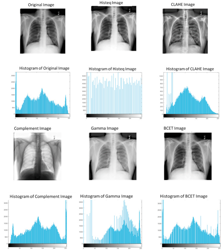
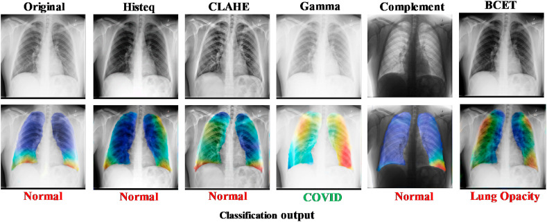
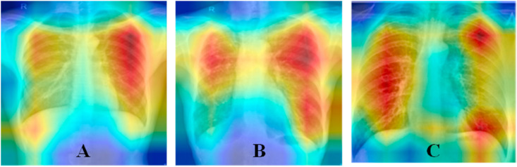
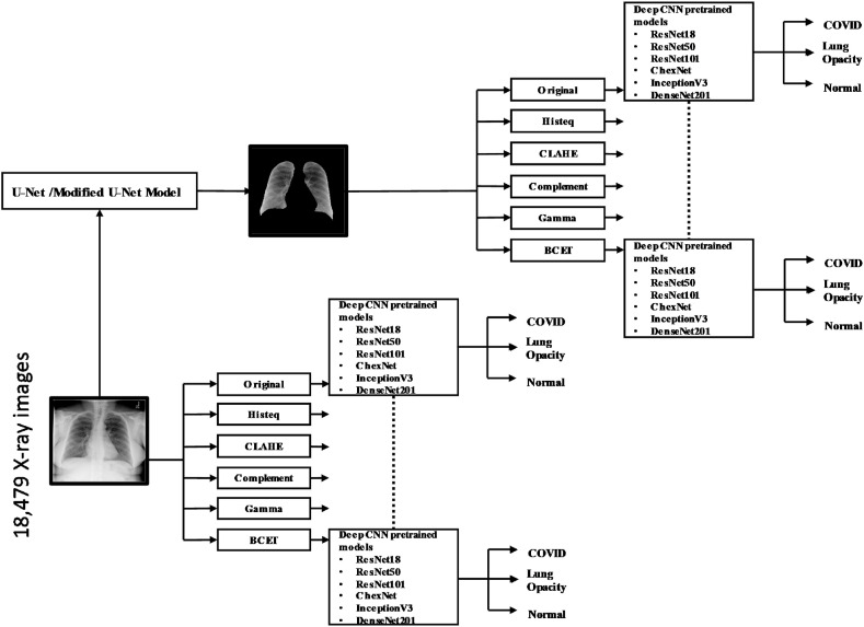
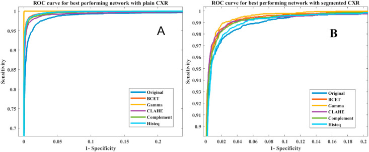
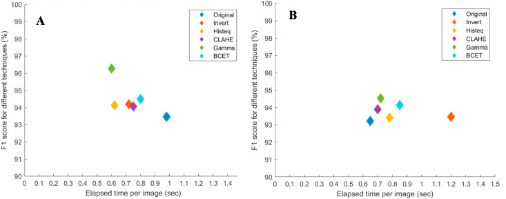
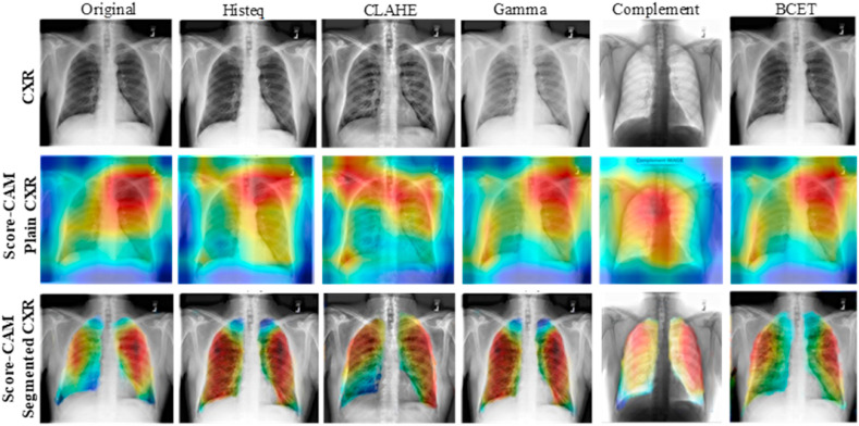
Computer-aided diagnosis for the reliable and fast detection of coronavirus disease (COVID-19) has become a necessity to prevent the spread of the virus during the pandemic to ease the burden on the healthcare system. Chest X-ray (CXR) imaging has several advantages over other imaging and detection techniques. Numerous works have been reported on COVID-19 detection from a smaller set of original X-ray images. However, the effect of image enhancement and lung segmentation of a large dataset in COVID-19 detection was not reported in the literature. We have compiled a large X-ray dataset (COVQU) consisting of 18,479 CXR images with 8851 normal, 6012 non-COVID lung infections, and 3616 COVID-19 CXR images and their corresponding ground truth lung masks. To the best of our knowledge, this is the largest public COVID positive database and the lung masks. Five different image enhancement techniques: histogram equalization (HE), contrast limited adaptive histogram equalization (CLAHE), image complement, gamma correction, and balance contrast enhancement technique (BCET) were used to investigate the effect of image enhancement techniques on COVID-19 detection. A novel U-Net model was proposed and compared with the standard U-Net model for lung segmentation. Six different pre-trained Convolutional Neural Networks (CNNs) (ResNet18, ResNet50, ResNet101, InceptionV3, DenseNet201, and ChexNet) and a shallow CNN model were investigated on the plain and segmented lung CXR images. The novel U-Net model showed an accuracy, Intersection over Union (IoU), and Dice coefficient of 98.63%, 94.3%, and 96.94%, respectively for lung segmentation. The gamma correction-based enhancement technique outperforms other techniques in detecting COVID-19 from the plain and the segmented lung CXR images. Classification performance from plain CXR images is slightly better than the segmented lung CXR images; however, the reliability of network performance is significantly improved for the segmented lung images, which was observed using the visualization technique. The accuracy, precision, sensitivity, F1-score, and specificity were 95.11%, 94.55%, 94.56%, 94.53%, and 95.59% respectively for the segmented lung images. The proposed approach with very reliable and comparable performance will boost the fast and robust COVID-19 detection using chest X-ray images.

摘要

计算机辅助诊断对于可靠和快速检测冠状病毒病(COVID-19)至关重要,可在大流行期间防止病毒传播,从而减轻医疗系统的负担。与其他成像和检测技术相比,胸部 X 射线(CXR)成像具有多种优势。已经有许多关于从较小的原始 X 射线图像集中检测 COVID-19 的工作。然而,在文献中并未报道在 COVID-19 检测中使用大量数据集进行图像增强和肺部分割的效果。我们已经编译了一个大型 X 射线数据集(COVQU),其中包含 18479 张 CXR 图像,其中 8851 张为正常,6012 张为非 COVID 肺部感染,3616 张为 COVID-19 CXR 图像及其相应的肺部蒙版。据我们所知,这是最大的公共 COVID 阳性数据库和肺部蒙版。我们使用了五种不同的图像增强技术:直方图均衡化(HE)、对比度限制自适应直方图均衡化(CLAHE)、图像补全、伽马校正和平衡对比度增强技术(BCET),以研究图像增强技术对 COVID-19 检测的影响。我们提出了一种新颖的 U-Net 模型,并将其与标准的 U-Net 模型进行了比较,用于肺部分割。我们研究了六种不同的预训练卷积神经网络(CNNs)(ResNet18、ResNet50、ResNet101、InceptionV3、DenseNet201 和 ChexNet)和一个浅层 CNN 模型在原始和分割的肺部 CXR 图像上的性能。新型 U-Net 模型的肺部分割准确性、交并比(IoU)和骰子系数分别为 98.63%、94.3%和 96.94%。基于伽马校正的增强技术在从原始和分割的肺部 CXR 图像中检测 COVID-19 方面优于其他技术。来自原始 CXR 图像的分类性能略优于分割的肺部 CXR 图像;然而,对于分割的肺部图像,网络性能的可靠性显著提高,这是通过可视化技术观察到的。对于分割的肺部图像,其准确性、精度、灵敏度、F1 分数和特异性分别为 95.11%、94.55%、94.56%、94.53%和 95.59%。我们提出的方法具有非常可靠和可比的性能,将促进使用胸部 X 射线图像进行快速和稳健的 COVID-19 检测。

https://cdn.ncbi.nlm.nih.gov/pmc/blobs/7056/7946571/d3edbea6c3ec/gr11_lrg.jpg
https://cdn.ncbi.nlm.nih.gov/pmc/blobs/7056/7946571/9d99e8b63818/gr1_lrg.jpg
https://cdn.ncbi.nlm.nih.gov/pmc/blobs/7056/7946571/e81fe35cfa6d/gr2_lrg.jpg
https://cdn.ncbi.nlm.nih.gov/pmc/blobs/7056/7946571/d8fa14360843/gr3_lrg.jpg
https://cdn.ncbi.nlm.nih.gov/pmc/blobs/7056/7946571/1e6628bf298b/gr4_lrg.jpg
https://cdn.ncbi.nlm.nih.gov/pmc/blobs/7056/7946571/496c5dc7e0e1/gr5_lrg.jpg
https://cdn.ncbi.nlm.nih.gov/pmc/blobs/7056/7946571/626b5719b4a6/gr6_lrg.jpg
https://cdn.ncbi.nlm.nih.gov/pmc/blobs/7056/7946571/50df1f0f48c9/gr7_lrg.jpg
https://cdn.ncbi.nlm.nih.gov/pmc/blobs/7056/7946571/b94e42d56f7e/gr8_lrg.jpg
https://cdn.ncbi.nlm.nih.gov/pmc/blobs/7056/7946571/5fc146182931/gr9_lrg.jpg
https://cdn.ncbi.nlm.nih.gov/pmc/blobs/7056/7946571/1dee2389ccc8/gr10_lrg.jpg
https://cdn.ncbi.nlm.nih.gov/pmc/blobs/7056/7946571/d3edbea6c3ec/gr11_lrg.jpg

相似文献

1
Exploring the effect of image enhancement techniques on COVID-19 detection using chest X-ray images.探讨使用胸部 X 光图像的图像增强技术对 COVID-19 检测的影响。
Comput Biol Med. 2021 May;132:104319. doi: 10.1016/j.compbiomed.2021.104319. Epub 2021 Mar 11.
2
Deep Learning for Reliable Classification of COVID-19, MERS, and SARS from Chest X-ray Images.基于胸部X光图像的深度学习对COVID-19、中东呼吸综合征和严重急性呼吸综合征进行可靠分类
Cognit Comput. 2022;14(5):1752-1772. doi: 10.1007/s12559-021-09955-1. Epub 2022 Jan 11.
3
COVID-19 infection localization and severity grading from chest X-ray images.通过胸部X光图像进行COVID-19感染定位及严重程度分级
Comput Biol Med. 2021 Dec;139:105002. doi: 10.1016/j.compbiomed.2021.105002. Epub 2021 Oct 30.
4
Optimized chest X-ray image semantic segmentation networks for COVID-19 early detection.用于 COVID-19 早期检测的优化胸部 X 射线图像语义分割网络。
J Xray Sci Technol. 2022;30(3):491-512. doi: 10.3233/XST-211113.
5
SVD-CLAHE boosting and balanced loss function for Covid-19 detection from an imbalanced Chest X-Ray dataset.SVD-CLAHE 增强和平衡损失函数在不平衡的 Chest X-Ray 数据集上进行 Covid-19 检测。
Comput Biol Med. 2022 Nov;150:106092. doi: 10.1016/j.compbiomed.2022.106092. Epub 2022 Sep 28.
6
Automatic coronavirus disease 2019 diagnosis based on chest radiography and deep learning - Success story or dataset bias?基于胸部 X 光和深度学习的新型冠状病毒病 2019 自动诊断——成功案例还是数据集偏差?
Med Phys. 2022 Feb;49(2):978-987. doi: 10.1002/mp.15419. Epub 2022 Jan 12.
7
LCSB-inception: Reliable and effective light-chroma separated branches for Covid-19 detection from chest X-ray images.LCSB-inception:从胸部 X 光图像中可靠且有效地检测新冠病毒的光-色分离分支。
Comput Biol Med. 2022 Nov;150:106195. doi: 10.1016/j.compbiomed.2022.106195. Epub 2022 Oct 14.
8
Chest X-ray image phase features for improved diagnosis of COVID-19 using convolutional neural network.基于卷积神经网络的胸部 X 射线图像相位特征提高 COVID-19 诊断性能
Int J Comput Assist Radiol Surg. 2021 Feb;16(2):197-206. doi: 10.1007/s11548-020-02305-w. Epub 2021 Jan 9.
9
Impact of Lung Segmentation on the Diagnosis and Explanation of COVID-19 in Chest X-ray Images.肺部分割对胸部 X 光图像中 COVID-19 诊断和解释的影响。
Sensors (Basel). 2021 Oct 27;21(21):7116. doi: 10.3390/s21217116.
10
Classification of COVID-19 chest X-Ray and CT images using a type of dynamic CNN modification method.利用一种动态卷积神经网络改进方法对 COVID-19 胸部 X 射线和 CT 图像进行分类。
Comput Biol Med. 2021 Jul;134:104425. doi: 10.1016/j.compbiomed.2021.104425. Epub 2021 Apr 29.

引用本文的文献

1
Segmentation-Assisted Fusion-Based Classification for Automated CXR Image Analysis.基于分割辅助融合的自动胸部X光图像分析分类方法
Sensors (Basel). 2025 Jul 24;25(15):4580. doi: 10.3390/s25154580.
2
Enhancing image retrieval through optimal barcode representation.通过优化条形码表示增强图像检索。
Sci Rep. 2025 Aug 7;15(1):28847. doi: 10.1038/s41598-025-14576-x.
3
A narrative review of foundation models for medical image segmentation: zero-shot performance evaluation on diverse modalities.医学图像分割基础模型的叙述性综述:不同模态下的零样本性能评估

本文引用的文献

1
Deep learning based detection and analysis of COVID-19 on chest X-ray images.基于深度学习的胸部X光图像中新型冠状病毒肺炎的检测与分析
Appl Intell (Dordr). 2021;51(3):1690-1700. doi: 10.1007/s10489-020-01902-1. Epub 2020 Oct 9.
2
Classification of COVID-19 in chest X-ray images using DeTraC deep convolutional neural network.使用DeTraC深度卷积神经网络对胸部X光图像中的新冠肺炎进行分类。
Appl Intell (Dordr). 2021;51(2):854-864. doi: 10.1007/s10489-020-01829-7. Epub 2020 Sep 5.
3
GraphXCOVID: Explainable deep graph diffusion pseudo-Labelling for identifying COVID-19 on chest X-rays.
Quant Imaging Med Surg. 2025 Jun 6;15(6):5825-5858. doi: 10.21037/qims-2024-2826. Epub 2025 Jun 3.
4
Gradual poisoning of a chest x-ray convolutional neural network with an adversarial attack and AI explainability methods.通过对抗攻击和人工智能可解释性方法对胸部X光卷积神经网络进行渐进式中毒攻击。
Sci Rep. 2025 Jul 1;15(1):21779. doi: 10.1038/s41598-025-02294-3.
5
Lessons learned from RadiologyNET foundation models for transfer learning in medical radiology.从放射学NET基础模型中汲取的经验教训,用于医学放射学中的迁移学习。
Sci Rep. 2025 Jul 1;15(1):21622. doi: 10.1038/s41598-025-05009-w.
6
A novel normalization algorithm to facilitate pre-assessment of Covid-19 disease by improving accuracy of CNN and its FPGA implementation.一种通过提高卷积神经网络(CNN)的准确性来促进新冠病毒疾病预评估的新型归一化算法及其现场可编程门阵列(FPGA)实现。
Evol Syst (Berl). 2022 Feb 1:1-11. doi: 10.1007/s12530-022-09419-3.
7
A multi-output network with U-net enhanced class activation map and robust classification performance for medical imaging analysis.一种用于医学影像分析的具有U型网络增强类激活映射和强大分类性能的多输出网络。
Discov Artif Intell. 2023;3(1):1. doi: 10.1007/s44163-022-00045-1. Epub 2023 Jan 3.
8
Deep viewing for the identification of Covid-19 infection status from chest X-Ray image using CNN based architecture.基于卷积神经网络架构,通过深度观察从胸部X光图像识别新冠病毒感染状态。
Intell Syst Appl. 2022 Nov;16:200130. doi: 10.1016/j.iswa.2022.200130. Epub 2022 Oct 6.
9
Sensitivity-Aware Differential Privacy for Federated Medical Imaging.用于联合医学成像的灵敏度感知差分隐私
Sensors (Basel). 2025 Apr 30;25(9):2847. doi: 10.3390/s25092847.
10
Unity in Diversity: Collaborative Pre-training Across Multimodal Medical Sources.多元中的统一:跨多模态医学资源的协作式预训练
Proc Conf Assoc Comput Linguist Meet. 2024 Aug;2024(Volume 1 Long Papers):3644-3656. doi: 10.18653/v1/2024.acl-long.199.
GraphXCOVID:用于在胸部X光片上识别新冠肺炎的可解释深度图扩散伪标签
Pattern Recognit. 2022 Feb;122:108274. doi: 10.1016/j.patcog.2021.108274. Epub 2021 Aug 26.
4
COVID-19 classification of X-ray images using deep neural networks.基于深度神经网络的 X 射线图像 COVID-19 分类。
Eur Radiol. 2021 Dec;31(12):9654-9663. doi: 10.1007/s00330-021-08050-1. Epub 2021 May 29.
5
RANDGAN: Randomized generative adversarial network for detection of COVID-19 in chest X-ray.RANDGAN:用于胸部 X 光 COVID-19 检测的随机生成对抗网络。
Sci Rep. 2021 Apr 21;11(1):8602. doi: 10.1038/s41598-021-87994-2.
6
Convolutional Sparse Support Estimator-Based COVID-19 Recognition From X-Ray Images.基于卷积稀疏支持估计器的 X 射线图像 COVID-19 识别
IEEE Trans Neural Netw Learn Syst. 2021 May;32(5):1810-1820. doi: 10.1109/TNNLS.2021.3070467. Epub 2021 May 3.
7
COVID-19 infection map generation and detection from chest X-ray images.从胸部X光图像生成并检测COVID-19感染地图。
Health Inf Sci Syst. 2021 Apr 1;9(1):15. doi: 10.1007/s13755-021-00146-8. eCollection 2021 Dec.
8
Thin-Section Chest CT Imaging of COVID-19 Pneumonia: A Comparison Between Patients with Mild and Severe Disease.新型冠状病毒肺炎的胸部薄层CT成像:轻型与重型患者的比较
Radiol Cardiothorac Imaging. 2020 Apr 23;2(2):e200126. doi: 10.1148/ryct.2020200126. eCollection 2020 Apr.
9
COVID-Net: a tailored deep convolutional neural network design for detection of COVID-19 cases from chest X-ray images.COVID-Net:一种针对胸部 X 光图像中 COVID-19 病例检测的定制化深度卷积神经网络设计。
Sci Rep. 2020 Nov 11;10(1):19549. doi: 10.1038/s41598-020-76550-z.
10
COVID-CAPS: A capsule network-based framework for identification of COVID-19 cases from X-ray images.COVID-CAPS:一种基于胶囊网络的从X射线图像识别新冠肺炎病例的框架。
Pattern Recognit Lett. 2020 Oct;138:638-643. doi: 10.1016/j.patrec.2020.09.010. Epub 2020 Sep 16.