Center for Gastrointestinal Research, Baylor Scott & White Research Institute and Charles A. Sammons Cancer Center, Baylor University Medical Center, Dallas, Texas; Department of Surgery, Tokushima University, Tokushima, Japan; Department of Molecular Diagnostics and Experimental Therapeutics, Beckman Research Institute of City of Hope Comprehensive Cancer Center, Duarte, California.
Department of Surgery, Tokushima University, Tokushima, Japan.
Gastroenterology. 2021 Jul;161(1):151-162.e1. doi: 10.1053/j.gastro.2021.03.062. Epub 2021 Apr 2.
BACKGROUND & AIMS: We recently reported use of tissue-based transcriptomic biomarkers (microRNA [miRNA] or messenger RNA [mRNA]) for identification of lymph node metastasis (LNM) in patients with invasive submucosal colorectal cancers (T1 CRC). In this study, we translated our tissue-based biomarkers into a blood-based liquid biopsy assay for noninvasive detection of LNM in patients with high-risk T1 CRC.
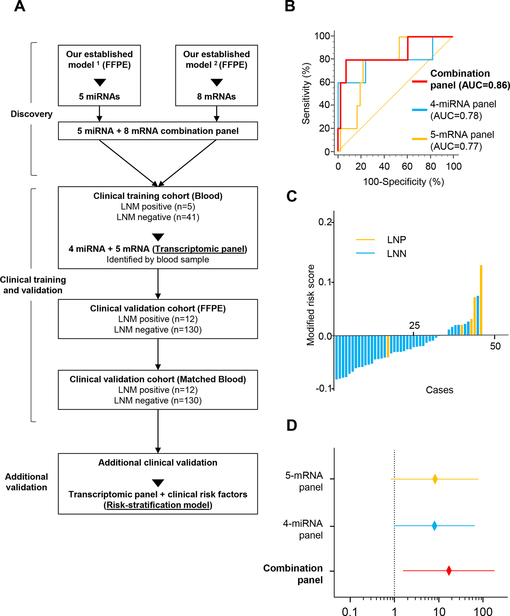
We analyzed 330 specimens from patients with high-risk T1 CRC, which included 188 serum samples from 2 clinical cohorts-a training cohort (N = 46) and a validation cohort (N = 142)-and matched formalin-fixed paraffin-embedded samples (N = 142). We performed quantitative reverse-transcription polymerase chain reaction, followed by logistic regression analysis, to develop an integrated transcriptomic panel and establish a risk-stratification model combined with clinical risk factors.
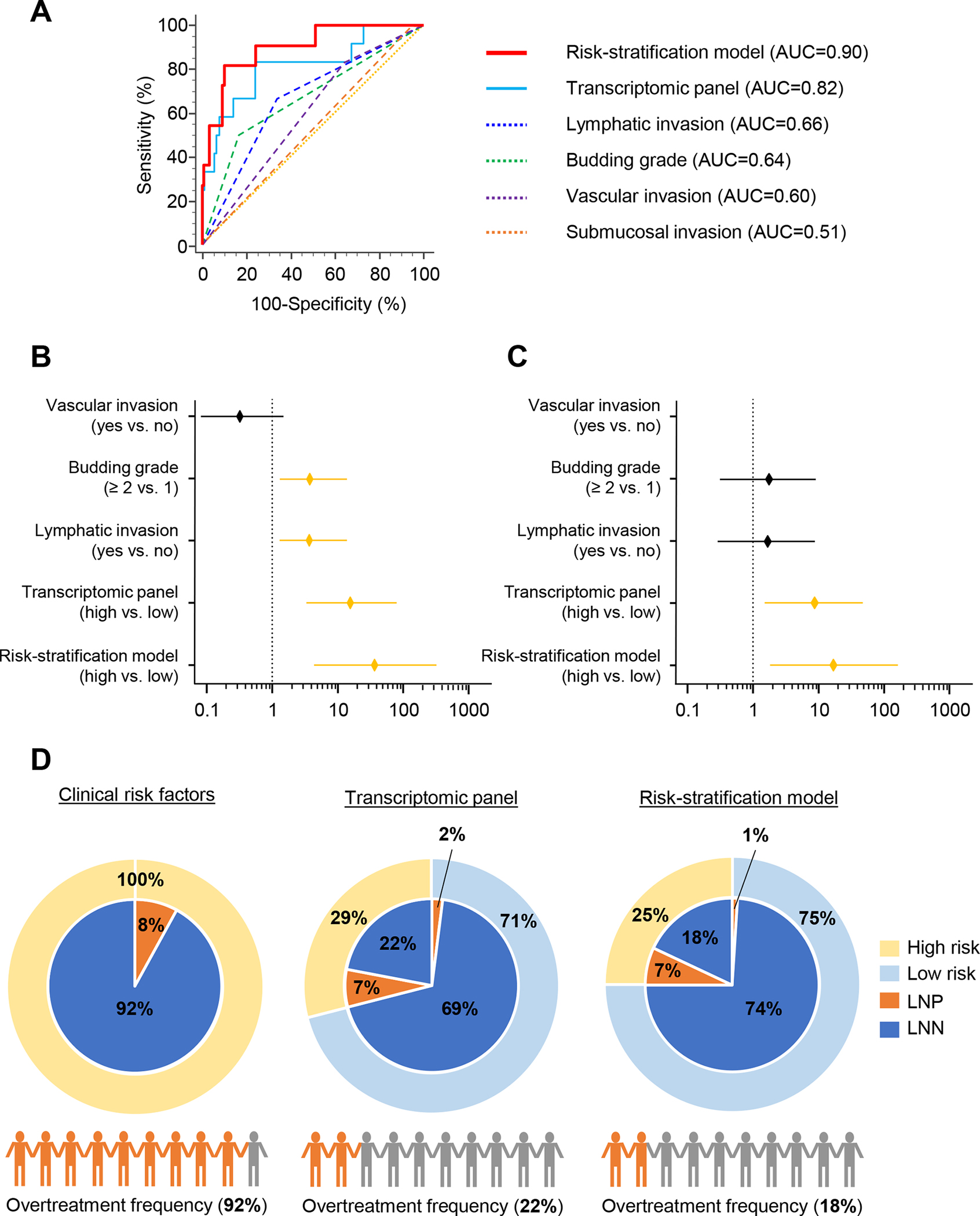
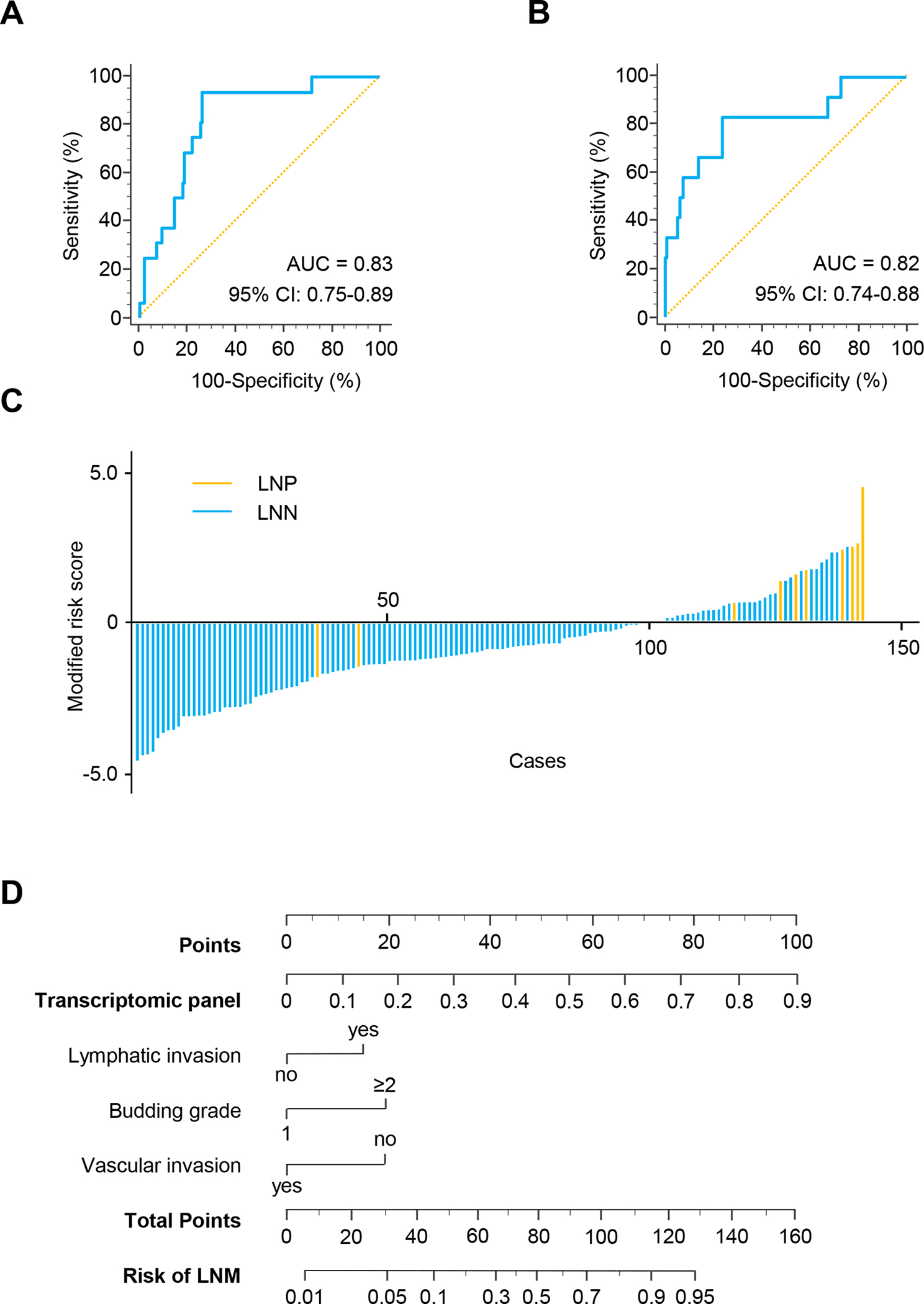
We used comprehensive expression profiling of a training cohort of LNM-positive and LMN-negative serum specimens to identify an optimized transcriptomic panel of 4 miRNAs (miR-181b, miR-193b, miR-195, and miR-411) and 5 mRNAs (AMT, forkhead box A1 [FOXA1], polymeric immunoglobulin receptor [PIGR], matrix metalloproteinase 1 [MMP1], and matrix metalloproteinase 9 [MMP9]), which robustly identified patients with LNM (area under the curve [AUC], 0.86; 95% confidence interval [CI], 0.72-0.94). We validated panel performance in an independent validation cohort (AUC, 0.82; 95% CI, 0.74-0.88). Our risk-stratification model was more accurate than the panel and an independent predictor for identification of LNM (AUC, 0.90; univariate: odds ratio [OR], 37.17; 95% CI, 4.48-308.35; P < .001; multivariate: OR, 17.28; 95% CI, 1.82-164.07; P = .013). The model limited potential overtreatment to only 18% of all patients, which is dramatically superior to pathologic features that are currently used (92%).
A novel risk-stratification model for noninvasive identification of T1 CRC has the potential to avoid unnecessary operations for patients classified as high-risk by conventional risk-classification criteria.
我们最近报道了使用基于组织的转录组生物标志物(microRNA [miRNA]或信使 RNA [mRNA])来识别浸润性黏膜下结直肠癌(T1 CRC)患者的淋巴结转移(LNM)。在这项研究中,我们将基于组织的生物标志物转化为一种基于血液的液体活检检测方法,用于非侵入性检测高危 T1 CRC 患者的 LNM。
我们分析了 330 份高危 T1 CRC 患者的标本,其中包括 2 个临床队列的 188 份血清样本(训练队列[N=46]和验证队列[N=142])和匹配的福尔马林固定石蜡包埋样本(N=142)。我们进行了定量逆转录聚合酶链反应,然后进行逻辑回归分析,以开发一个综合转录组面板,并建立一个结合临床危险因素的风险分层模型。
我们使用 LNM 阳性和 LNM 阴性血清标本的训练队列进行全面表达谱分析,确定了一个由 4 个 miRNA(miR-181b、miR-193b、miR-195 和 miR-411)和 5 个 mRNA(AMT、叉头框 A1 [FOXA1]、多聚免疫球蛋白受体 [PIGR]、基质金属蛋白酶 1 [MMP1]和基质金属蛋白酶 9 [MMP9])组成的优化转录组面板,该面板可稳健地识别 LNM 患者(曲线下面积[AUC],0.86;95%置信区间[CI],0.72-0.94)。我们在独立验证队列中验证了该面板的性能(AUC,0.82;95%CI,0.74-0.88)。我们的风险分层模型比面板和独立预测因子更准确,用于识别 LNM(AUC,0.90;单变量:优势比[OR],37.17;95%CI,4.48-308.35;P<0.001;多变量:OR,17.28;95%CI,1.82-164.07;P=0.013)。该模型将潜在的过度治疗仅限于所有患者的 18%,这明显优于目前使用的病理特征(92%)。
一种用于非侵入性识别 T1 CRC 的新型风险分层模型有可能避免对传统风险分类标准分类为高危的患者进行不必要的手术。