State Key Laboratory of Molecular Biology, Shanghai Key Laboratory of Molecular Andrology, CAS Center for Excellence in Molecular Cell Science, Shanghai Institute of Biochemistry and Cell Biology, University of Chinese Academy of Sciences, Chinese Academy of Sciences, Shanghai 200031, China.
School of Life Science and Technology, ShanghaiTech University, Shanghai 201210, China.
RNA. 2021 Jun;27(6):725-733. doi: 10.1261/rna.078671.120. Epub 2021 Apr 12.
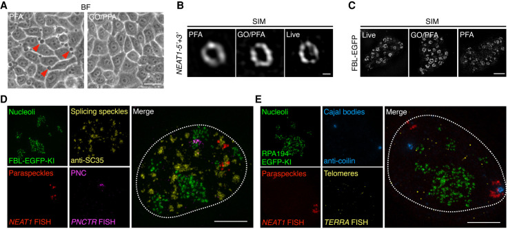
The mammalian cell nucleus contains different types of membrane-less nuclear bodies (NBs) consisting of proteins and RNAs. Microscopic imaging has been widely applied to study the organization and structure of NBs. However, current fixation methods are not optimized for such imaging: When a fixation method is chosen to maximize the quality of the RNA fluorescence in situ hybridization (FISH), it often limits the labeling efficiency of proteins or affects the ultrastructure of NBs. Here, we report that addition of glyoxal (GO) into the classical paraformaldehyde (PFA) fixation step not only improves FISH signals for RNAs in NBs via augmented permeability of the fixed nucleus and enhanced accessibility of probes, but also largely preserves protein fluorescent signals during fixation and immunostaining. We also show that GO/PFA fixation enables the covisualization of different types of nuclear bodies with minimal impact on their ultrastructures under super-resolution microscopy.
哺乳动物细胞核包含不同类型的无膜核体(NBs),由蛋白质和 RNA 组成。显微镜成像已广泛应用于研究 NBs 的组织和结构。然而,目前的固定方法并不针对这种成像进行优化:当选择一种固定方法以使 RNA 荧光原位杂交(FISH)的质量最大化时,它通常会限制蛋白质的标记效率或影响 NBs 的超微结构。在这里,我们报告称,在经典的多聚甲醛(PFA)固定步骤中加入乙二醛(GO)不仅通过增加固定核的通透性和增强探针的可及性来提高 NBs 中 RNA 的 FISH 信号,而且在固定和免疫染色过程中还能很大程度上保留蛋白质荧光信号。我们还表明,GO/PFA 固定在超分辨率显微镜下最小化对不同类型核体的超微结构的影响,从而实现不同类型核体的共可视化。