Calu Valentin, Ionescu Adriana, Stanca Loredana, Geicu Ovidiu Ionut, Iordache Florin, Pisoschi Aurelia Magdalena, Serban Andreea Iren, Bilteanu Liviu
Department of General Surgery, University of Medicine and Pharmacy "Carol Davila" Bucharest, 8 Blvd., Eroii Sanitari, 050474, Bucharest, Romania.
Department of Surgery, "Elias" Emergency University Hospital, 17 Marasti Blvd., 01146, Bucharest, Romania.
Sci Rep. 2021 Apr 12;11(1):7940. doi: 10.1038/s41598-021-86941-5.
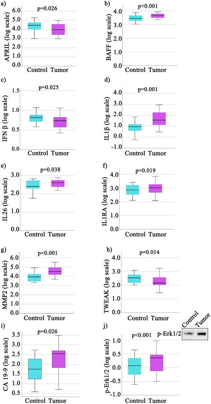
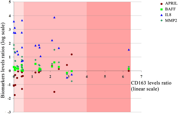
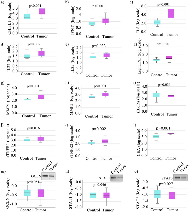
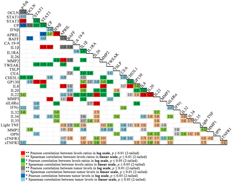
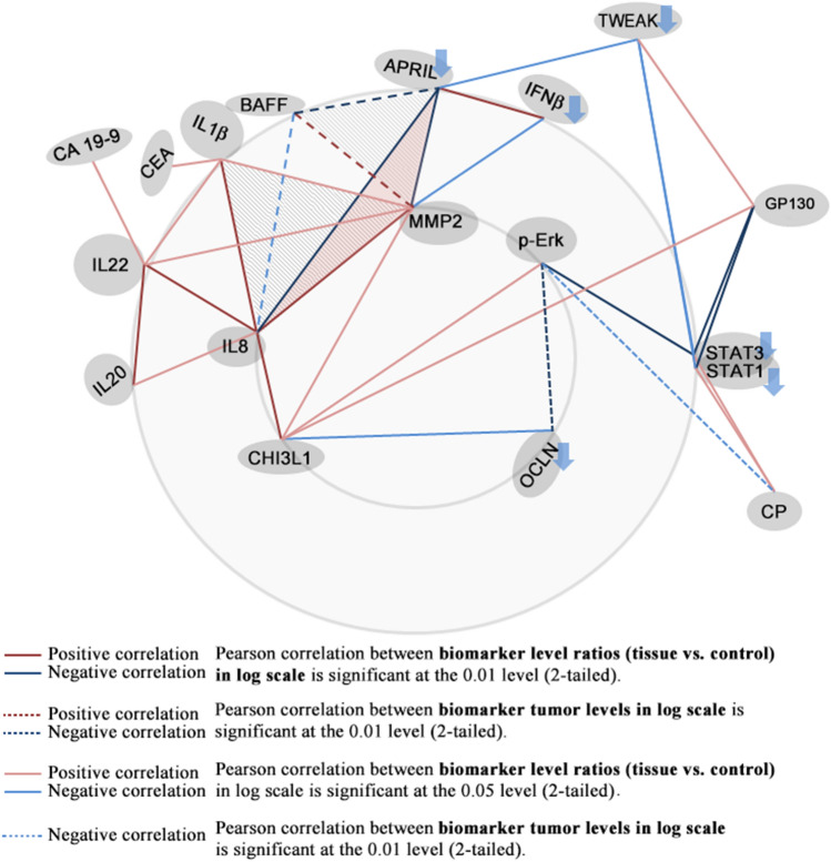
Therapeutic approaches focused on the inflammatory microenvironment are currently gaining more support, as biomolecules involved in the inflammatory colorectal cancer (CRC) tumor microenvironment are being explored. We analyzed tumor and paired normal tissue samples from CRC patients (n = 22) whom underwent tumor resection surgery. We assessed 39 inflammation-involved biomolecules (multiplex magnetic bead-based immunoassay), CEA and CA19-9 (ELISA assay) and the tissue expression levels of occludin and also pErk, STAT1 and STAT3 transcriptional factors (western blot). Tumor staging has been established by histopathological evaluation of HE stained tumor tissue sections. We report 32 biomarkers displaying statistically significant differences in tumor vs. control. Additionally, positive statistical biomarker correlations were found between MMP2-IL8 and BAFF-IL8 (Pearson correlation coefficients > 0.751), while APRIL-MMP2, APRIL-BAFF and APRIL-IL8 were negatively correlated (correlation coefficients < - 0.650). While APRIL, BAFF, IL8 and MMP2 did not modulate with tumor stage, they were inversely related to the immune infiltrate level and CD163 tissue expression. We conclude that the significantly decreased APRIL and increased BAFF, IL8 and MMP2 expression were tumor-specific and deserve consideration in the development of new treatments. Also, the positive correlation between Chitinase 3-like 1 and IL8 (0.57) or MMP2 (0.50) suggest a role in tumor growth and metastasis pathways.
随着参与炎症性结直肠癌(CRC)肿瘤微环境的生物分子不断被探索,聚焦于炎症微环境的治疗方法目前正获得更多支持。我们分析了22例接受肿瘤切除手术的CRC患者的肿瘤组织及配对的正常组织样本。我们评估了39种参与炎症的生物分子(基于多重磁珠的免疫测定法)、癌胚抗原(CEA)和糖类抗原19-9(CA19-9,酶联免疫吸附测定法),以及闭合蛋白、磷酸化细胞外信号调节激酶(pErk)、信号转导和转录激活因子1(STAT1)和信号转导和转录激活因子3(STAT3)转录因子的组织表达水平(蛋白质免疫印迹法)。通过对苏木精-伊红(HE)染色的肿瘤组织切片进行组织病理学评估来确定肿瘤分期。我们报告了32种生物标志物在肿瘤组织与对照组织中显示出统计学上的显著差异。此外,基质金属蛋白酶2(MMP2)与白细胞介素8(IL8)、B细胞活化因子(BAFF)与IL8之间存在正相关的统计学生物标志物关联(皮尔逊相关系数>0.751),而增殖诱导配体(APRIL)与MMP2、APRIL与BAFF以及APRIL与IL8呈负相关(相关系数<-0.650)。虽然APRIL、BAFF、IL8和MMP2不随肿瘤分期而调节,但它们与免疫浸润水平和CD163组织表达呈负相关。我们得出结论,APRIL显著降低以及BAFF、IL8和MMP2表达增加具有肿瘤特异性,值得在开发新治疗方法时予以考虑。此外,几丁质酶3样1与IL8(0.57)或MMP2(0.50)之间的正相关表明其在肿瘤生长和转移途径中发挥作用。