Alphandéry Edouard
Paris Sorbonne Université, Muséum National d'Histoire Naturelle, UMR CNRS7590, IRD, Institut de Minéralogie, de Physique des Matériaux et deCosmochimie, IMPMC 75005 Paris France.
Nanobacterie SARL 36 Boulevard Flandrin 75116 Paris France.
RSC Adv. 2019 Dec 6;9(69):40577-40587. doi: 10.1039/c9ra08612a. eCollection 2019 Dec 3.
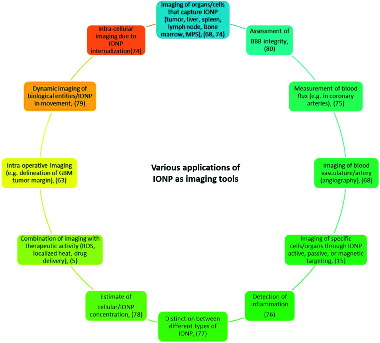
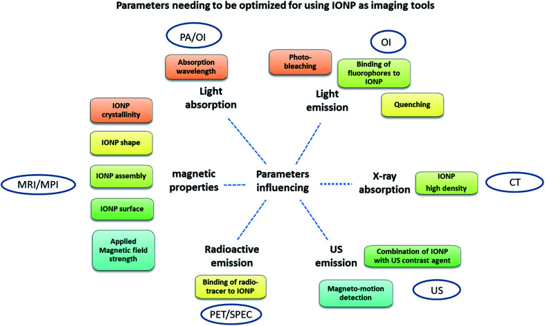
In medicine, obtaining simply a resolute and accurate image of an organ of interest is a real challenge. To achieve this, it has recently been proposed to use combined methods in which standard imaging (MRI, PAI, CT, PET/SPEC, USI, OI) is carried out in the presence of iron oxide nanoparticles, thus making it possible to image certain tissues/cells through the specific targeting of these nanoparticles, hence resulting in improved imaging contrast and resolution. Here, the advantages and drawbacks of these combined methods are presented as well as some of their recent medical applications.
在医学领域,仅仅获得感兴趣器官的清晰准确图像是一项真正的挑战。为实现这一目标,最近有人提出使用联合方法,即在存在氧化铁纳米颗粒的情况下进行标准成像(磁共振成像、光声成像、计算机断层扫描、正电子发射断层扫描/单光子发射计算机断层扫描、超声成像、光学成像),从而能够通过这些纳米颗粒的特异性靶向对某些组织/细胞进行成像,进而提高成像对比度和分辨率。本文介绍了这些联合方法的优缺点及其一些近期的医学应用。