Cheng Jun, Liu Yuting, Huang Wei, Hong Wenhui, Wang Lingling, Zhan Xiaohui, Han Zhi, Ni Dong, Huang Kun, Zhang Jie
National-Regional Key Technology Engineering Laboratory for Medical Ultrasound, Shenzhen University, Shenzhen, China.
Guangdong Key Laboratory for Biomedical Measurements and Ultrasound Imaging, School of Biomedical Engineering, Health Science Center, Shenzhen University, Shenzhen, China.
Front Oncol. 2021 Mar 31;11:623382. doi: 10.3389/fonc.2021.623382. eCollection 2021.
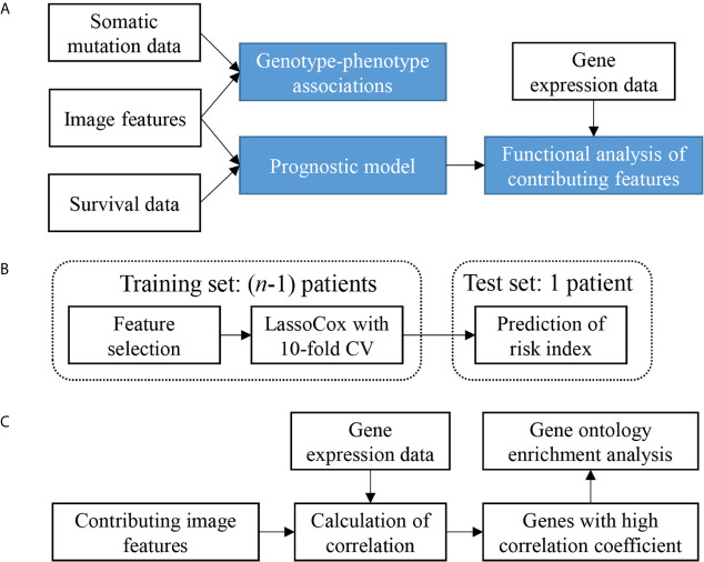
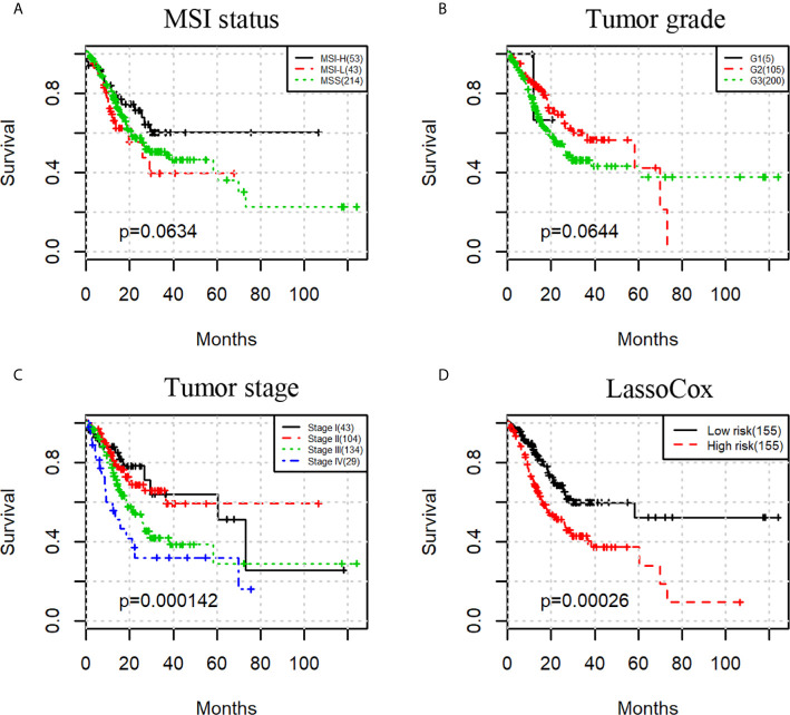
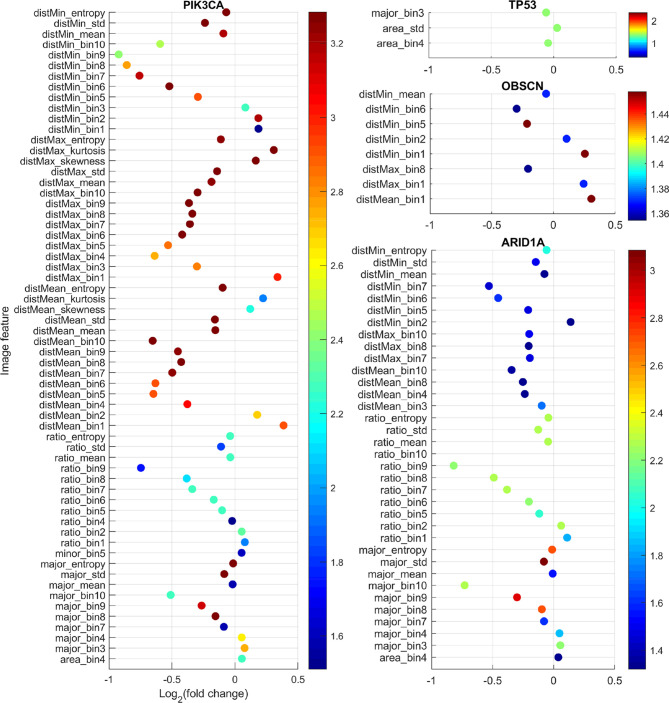
Computational analysis of histopathological images can identify sub-visual objective image features that may not be visually distinguishable by human eyes, and hence provides better modeling of disease phenotypes. This study aims to investigate whether specific image features are associated with somatic mutations and patient survival in gastric adenocarcinoma (sample size = 310). An automated image analysis pipeline was developed to extract quantitative morphological features from H&E stained whole-slide images. We found that four frequently somatically mutated genes (TP53, ARID1A, OBSCN, and PIK3CA) were significantly associated with tumor morphological changes. A prognostic model built on the image features significantly stratified patients into low-risk and high-risk groups (log-rank test p-value = 2.6e-4). Multivariable Cox regression showed the model predicted risk index was an additional prognostic factor besides tumor grade and stage. Gene ontology enrichment analysis showed that the genes whose expressions mostly correlated with the contributing features in the prognostic model were enriched on biological processes such as cell cycle and muscle contraction. These results demonstrate that histopathological image features can reflect underlying somatic mutations and identify high-risk patients that may benefit from more precise treatment regimens. Both the image features and pipeline are highly interpretable to enable translational applications.
组织病理学图像的计算分析能够识别肉眼可能无法视觉分辨的亚视觉客观图像特征,从而更好地对疾病表型进行建模。本研究旨在调查在胃腺癌中(样本量 = 310)特定图像特征是否与体细胞突变及患者生存率相关。开发了一种自动图像分析流程,以从苏木精 - 伊红(H&E)染色的全切片图像中提取定量形态特征。我们发现四个频繁发生体细胞突变的基因(TP53、ARID1A、OBSCN和PIK3CA)与肿瘤形态变化显著相关。基于图像特征构建的预后模型将患者显著分层为低风险和高风险组(对数秩检验p值 = 2.6e - 4)。多变量Cox回归显示,该模型预测的风险指数是除肿瘤分级和分期之外的另一个预后因素。基因本体富集分析表明,其表达与预后模型中贡献特征大多相关的基因在细胞周期和肌肉收缩等生物学过程中富集。这些结果表明,组织病理学图像特征可以反映潜在的体细胞突变,并识别可能从更精确治疗方案中受益的高风险患者。图像特征和流程都具有高度可解释性,以实现转化应用。