• 文献检索
  • 文档翻译
  • 深度研究
  • 学术资讯
  • Suppr Zotero 插件Zotero 插件
  • 邀请有礼
  • 套餐&价格
  • 历史记录
应用&插件
Suppr Zotero 插件Zotero 插件浏览器插件Mac 客户端Windows 客户端微信小程序
定价
高级版会员购买积分包购买API积分包
服务
文献检索文档翻译深度研究API 文档MCP 服务
关于我们
关于 Suppr公司介绍联系我们用户协议隐私条款
关注我们

Suppr 超能文献

核心技术专利:CN118964589B侵权必究
粤ICP备2023148730 号-1Suppr @ 2026

文献检索

告别复杂PubMed语法,用中文像聊天一样搜索,搜遍4000万医学文献。AI智能推荐,让科研检索更轻松。

立即免费搜索

文件翻译

保留排版,准确专业,支持PDF/Word/PPT等文件格式,支持 12+语言互译。

免费翻译文档

深度研究

AI帮你快速写综述,25分钟生成高质量综述,智能提取关键信息,辅助科研写作。

立即免费体验

正常眼压性青光眼和原发性开角型青光眼患者的视盘周围血管密度比较,这些患者的青光眼损害程度相似。

A comparison of peripapillary vessel density between subjects with normal-tension glaucoma and primary open-angle glaucoma with similar extents of glaucomatous damage.

机构信息

Department of Ophthalmology, Konyang University College of Medicine, Daejeon, Republic of Korea.

Modoo's Eye Clinic, #238, Daedeok-daero, Seo-gu, Daejeon, Republic of Korea.

出版信息

Sci Rep. 2023 Jun 7;13(1):9258. doi: 10.1038/s41598-023-36369-w.

DOI:10.1038/s41598-023-36369-w
PMID:37286792
原文链接:https://pmc.ncbi.nlm.nih.gov/articles/PMC10247707/
Abstract

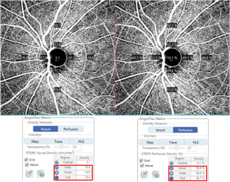
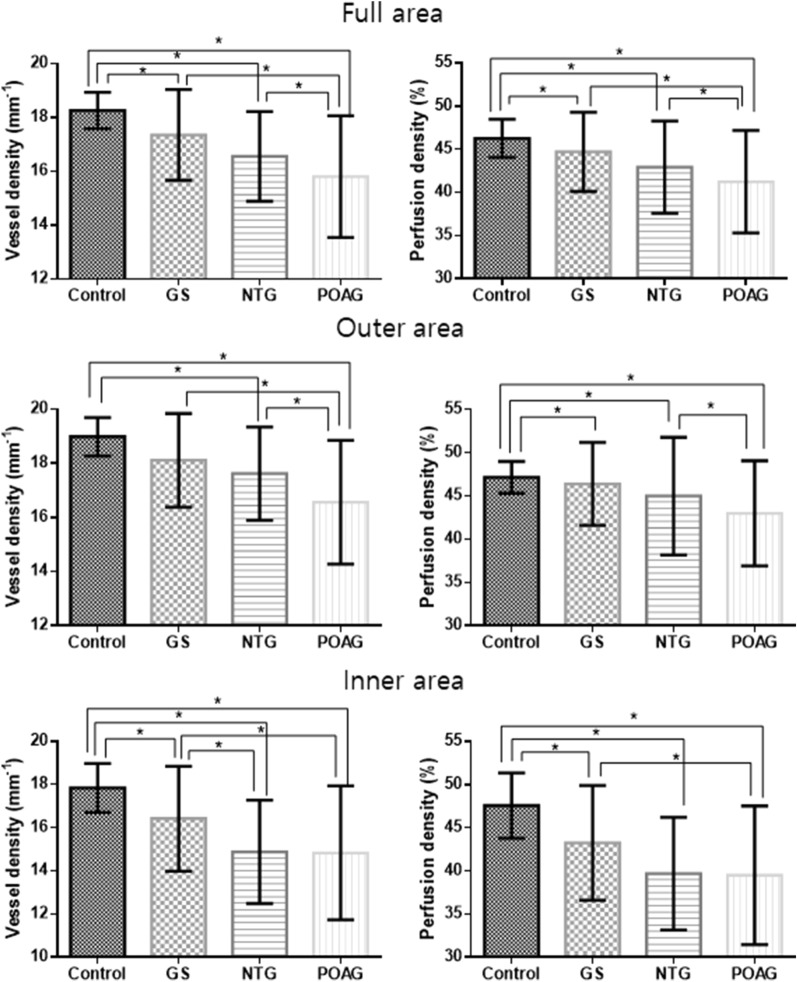
The purpose of this study was to identify differences in retinal microvasculature impairments between patients with normal-tension glaucoma (NTG) and those with primary open-angle glaucoma (POAG) with similar extents of structural and visual field damage. Participants with glaucoma-suspect (GS), NTG, POAG, and normal controls were consecutively enrolled. Peripapillary vessel density (VD) and perfusion density (PD) were compared among the groups. Linear regression analyses were performed to identify the relationship between VD, PD and visual field parameters. The VDs of the full areas were 18.3 ± 0.7, 17.3 ± 1.7, 16.5 ± 1.7, and 15.8 ± 2.3 mm in the control, GS, NTG, and POAG groups, respectively (P < 0.001). The VDs of the outer and inner areas and the PDs of all areas also differed significantly among the groups (all P < 0.001). In the NTG group, the VDs of the full, outer, and inner areas were significantly associated with all visual field parameters including the mean deviation (MD), pattern standard deviation (PSD), and visual field index (VFI). In the POAG group, the VDs of the full and inner areas were significantly associated with PSD and VFI but not with MD. In conclusion, with similar degrees of retinal nerve fiber layer thinning and visual field damage in both groups, the POAG group showed a lower peripapillary VD and PD than the NTG group. VD and PD were significantly associated with visual field loss.

摘要

本研究旨在确定具有相似程度的视网膜神经纤维层变薄和视野损伤的正常眼压性青光眼(NTG)和原发性开角型青光眼(POAG)患者之间视网膜微血管损伤的差异。连续纳入疑似青光眼(GS)、NTG、POAG 和正常对照组的患者。比较各组的视盘周围血管密度(VD)和灌注密度(PD)。进行线性回归分析以确定 VD、PD 与视野参数之间的关系。对照组、GS 组、NTG 组和 POAG 组的全区域 VD 分别为 18.3±0.7、17.3±1.7、16.5±1.7 和 15.8±2.3mm(P<0.001)。外区和内区的 VD 以及所有区域的 PD 在各组之间也有显著差异(均 P<0.001)。在 NTG 组中,全区域、外区域和内区域的 VD 与所有视野参数(包括平均偏差(MD)、模式标准差(PSD)和视野指数(VFI))均显著相关。在 POAG 组中,全区域和内区域的 VD 与 PSD 和 VFI 显著相关,但与 MD 无关。总之,在两组视网膜神经纤维层变薄和视野损伤程度相似的情况下,POAG 组的视盘周围 VD 和 PD 低于 NTG 组。VD 和 PD 与视野丧失显著相关。

https://cdn.ncbi.nlm.nih.gov/pmc/blobs/5c65/10247707/945b1d5f3f0f/41598_2023_36369_Fig2_HTML.jpg
https://cdn.ncbi.nlm.nih.gov/pmc/blobs/5c65/10247707/e190d9ede7f1/41598_2023_36369_Fig1_HTML.jpg
https://cdn.ncbi.nlm.nih.gov/pmc/blobs/5c65/10247707/945b1d5f3f0f/41598_2023_36369_Fig2_HTML.jpg
https://cdn.ncbi.nlm.nih.gov/pmc/blobs/5c65/10247707/e190d9ede7f1/41598_2023_36369_Fig1_HTML.jpg
https://cdn.ncbi.nlm.nih.gov/pmc/blobs/5c65/10247707/945b1d5f3f0f/41598_2023_36369_Fig2_HTML.jpg

相似文献

1
A comparison of peripapillary vessel density between subjects with normal-tension glaucoma and primary open-angle glaucoma with similar extents of glaucomatous damage.正常眼压性青光眼和原发性开角型青光眼患者的视盘周围血管密度比较,这些患者的青光眼损害程度相似。
Sci Rep. 2023 Jun 7;13(1):9258. doi: 10.1038/s41598-023-36369-w.
2
Quantitative Analysis of Microvasculature in Macular and Peripapillary Regions in Early Primary Open-Angle Glaucoma.早期原发性开角型青光眼黄斑及视盘周围区域微血管定量分析。
Curr Eye Res. 2020 May;45(5):629-635. doi: 10.1080/02713683.2019.1676912. Epub 2019 Oct 14.
3
Optical Coherence Tomography Angiography Analysis of Perfused Peripapillary Capillaries in Primary Open-Angle Glaucoma and Normal-Tension Glaucoma.原发性开角型青光眼和正常眼压性青光眼视乳头周围灌注毛细血管的光学相干断层扫描血管造影分析
Invest Ophthalmol Vis Sci. 2016 Jul 1;57(9):OCT611-OCT620. doi: 10.1167/iovs.15-18945.
4
Optic Disc Perfusion in Primary Open Angle and Normal Tension Glaucoma Eyes Using Optical Coherence Tomography-Based Microangiography.使用基于光学相干断层扫描的微血管造影术评估原发性开角型青光眼和正常眼压性青光眼患者视盘的血流灌注情况
PLoS One. 2016 May 5;11(5):e0154691. doi: 10.1371/journal.pone.0154691. eCollection 2016.
5
Topographic Differences in Superficial Macular Vessel Density in Eyes with Early Primary Open-Angle Glaucoma and Normal Tension Glaucoma.早期原发性开角型青光眼和正常眼压型青光眼患者浅层视网膜血管密度的地形差异。
Ophthalmic Res. 2023;66(1):465-473. doi: 10.1159/000528877. Epub 2023 Jan 5.
6
Optical Coherence Tomography Angiography of Optic Disc in Eyes With Primary Open-angle Glaucoma and Normal-tension Glaucoma.原发性开角型青光眼和正常眼压性青光眼患者视盘的光学相干断层扫描血管造影。
J Glaucoma. 2019 Mar;28(3):243-251. doi: 10.1097/IJG.0000000000001184.
7
Comparison of macular ganglion cell complex thickness by Fourier-domain OCT in normal tension glaucoma and primary open-angle glaucoma.正常眼压性青光眼和原发性开角型青光眼的频域光相干断层扫描测量的黄斑神经节细胞复合体厚度比较。
J Glaucoma. 2013 Feb;22(2):133-9. doi: 10.1097/IJG.0b013e3182254cde.
8
Comparison of microvascular parameters and diagnostic ability of optical coherence tomography angiography between eyes with primary angle closure glaucoma and primary open angle glaucoma.原发性闭角型青光眼与原发性开角型青光眼患者眼部的微血管参数及光学相干断层扫描血管造影诊断能力的比较
Photodiagnosis Photodyn Ther. 2022 Dec;40:103114. doi: 10.1016/j.pdpdt.2022.103114. Epub 2022 Sep 10.
9
Peripapillary and Macular Vessel Density Measurement by Optical Coherence Tomography Angiography in Pseudoexfoliation and Primary Open-angle Glaucoma.光学相干断层扫描血管造影术在假性剥脱综合征和原发性开角型青光眼的周边和黄斑血管密度测量。
J Glaucoma. 2020 May;29(5):381-385. doi: 10.1097/IJG.0000000000001464.
10
Comparison of retinal nerve fiber layer and macular thickness for discriminating primary open-angle glaucoma and normal-tension glaucoma using optical coherence tomography.使用光学相干断层扫描比较视网膜神经纤维层和黄斑厚度以鉴别原发性开角型青光眼和正常眼压性青光眼。
Clin Exp Optom. 2016 Jul;99(4):373-81. doi: 10.1111/cxo.12366. Epub 2016 Mar 21.

引用本文的文献

1
Peripapillary Perfusion Analysis Using Optical Coherence Tomography Angiography in Patients with Normal Tension Glaucoma.用光学相干断层扫描血管造影术分析正常眼压青光眼患者的视盘周围灌注。
Korean J Ophthalmol. 2024 Feb;38(1):51-63. doi: 10.3341/kjo.2023.0112. Epub 2023 Dec 19.
2
Vertical asymmetry analysis of the macular microvasculature in epiretinal membrane patients with open-angle glaucoma.特发性黄斑裂孔视网膜前膜患者伴开角型青光眼的黄斑微血管垂直不对称性分析。
Sci Rep. 2023 Oct 10;13(1):17053. doi: 10.1038/s41598-023-44053-2.

本文引用的文献

1
Change of Retinal Vessel Density After Lowering Intraocular Pressure in Ocular Hypertension.高眼压症患者降低眼压后视网膜血管密度的变化
Front Med (Lausanne). 2021 Dec 9;8:730327. doi: 10.3389/fmed.2021.730327. eCollection 2021.
2
Investigation of the Peripapillary Choriocapillaris in Normal Tension Glaucoma, Primary Open-angle Glaucoma, and Control Eyes.正常眼压性青光眼、原发性开角型青光眼和对照眼中的视盘周围脉络膜毛细血管研究。
J Glaucoma. 2021 Aug 1;30(8):682-689. doi: 10.1097/IJG.0000000000001861.
3
Relationship between retinal capillary vessel density of OCT angiography and intraocular pressure in pig.
猪眼 OCT 血管造影视网膜毛细血管密度与眼压的关系。
Sci Rep. 2021 Apr 20;11(1):8555. doi: 10.1038/s41598-021-87689-8.
4
Parafoveal vessel changes in primary open-angle glaucoma and normal-tension glaucoma using optical coherence tomography angiography.使用光学相干断层扫描血管造影术观察原发性开角型青光眼和正常眼压性青光眼的黄斑旁血管变化
Clin Ophthalmol. 2019 Sep 27;13:1935-1945. doi: 10.2147/OPTH.S206288. eCollection 2019.
5
Macula Vessel Density and Thickness in Early Primary Open-Angle Glaucoma.早期原发性开角型青光眼的黄斑血管密度和厚度。
Am J Ophthalmol. 2019 Mar;199:120-132. doi: 10.1016/j.ajo.2018.11.012. Epub 2018 Nov 26.
6
Optical Coherence Tomography Angiography Macular Vascular Density Measurements and the Central 10-2 Visual Field in Glaucoma.光学相干断层扫描血管造影黄斑血管密度测量与青光眼中心 10-2 视野。
J Glaucoma. 2018 Jun;27(6):481-489. doi: 10.1097/IJG.0000000000000964.
7
Inter-eye Asymmetry of Optical Coherence Tomography Angiography Vessel Density in Bilateral Glaucoma, Glaucoma Suspect, and Healthy Eyes.双眼光学相干断层扫描血管造影血管密度的不对称性在双侧青光眼、青光眼疑似患者和正常眼中的表现。
Am J Ophthalmol. 2018 Jun;190:69-77. doi: 10.1016/j.ajo.2018.03.026. Epub 2018 Mar 24.
8
Progressive Macula Vessel Density Loss in Primary Open-Angle Glaucoma: A Longitudinal Study.原发性开角型青光眼黄斑区血管密度的渐进性丧失:一项纵向研究。
Am J Ophthalmol. 2017 Oct;182:107-117. doi: 10.1016/j.ajo.2017.07.011. Epub 2017 Jul 20.
9
Regional vascular density-visual field sensitivity relationship in glaucoma according to disease severity.青光眼患者中根据疾病严重程度的局部血管密度与视野敏感度关系
Br J Ophthalmol. 2017 Dec;101(12):1666-1672. doi: 10.1136/bjophthalmol-2017-310180. Epub 2017 Apr 21.
10
Optical Coherence Tomography Angiography Analysis of Perfused Peripapillary Capillaries in Primary Open-Angle Glaucoma and Normal-Tension Glaucoma.原发性开角型青光眼和正常眼压性青光眼视乳头周围灌注毛细血管的光学相干断层扫描血管造影分析
Invest Ophthalmol Vis Sci. 2016 Jul 1;57(9):OCT611-OCT620. doi: 10.1167/iovs.15-18945.