McMahon Nathan P, Jones Jocelyn A, Anderson Ashley N, Dietz Matthew S, Wong Melissa H, Gibbs Summer L
Biomedical Engineering Department, Oregon Health & Science University, Portland, OR 97201, USA.
Department of Cell, Development & Cancer Biology Department, Oregon Health & Science University, Portland, OR 97201, USA.
Cancers (Basel). 2023 Jan 29;15(3):827. doi: 10.3390/cancers15030827.
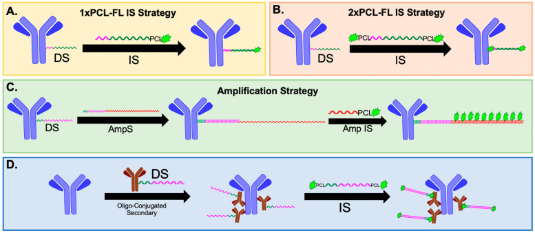
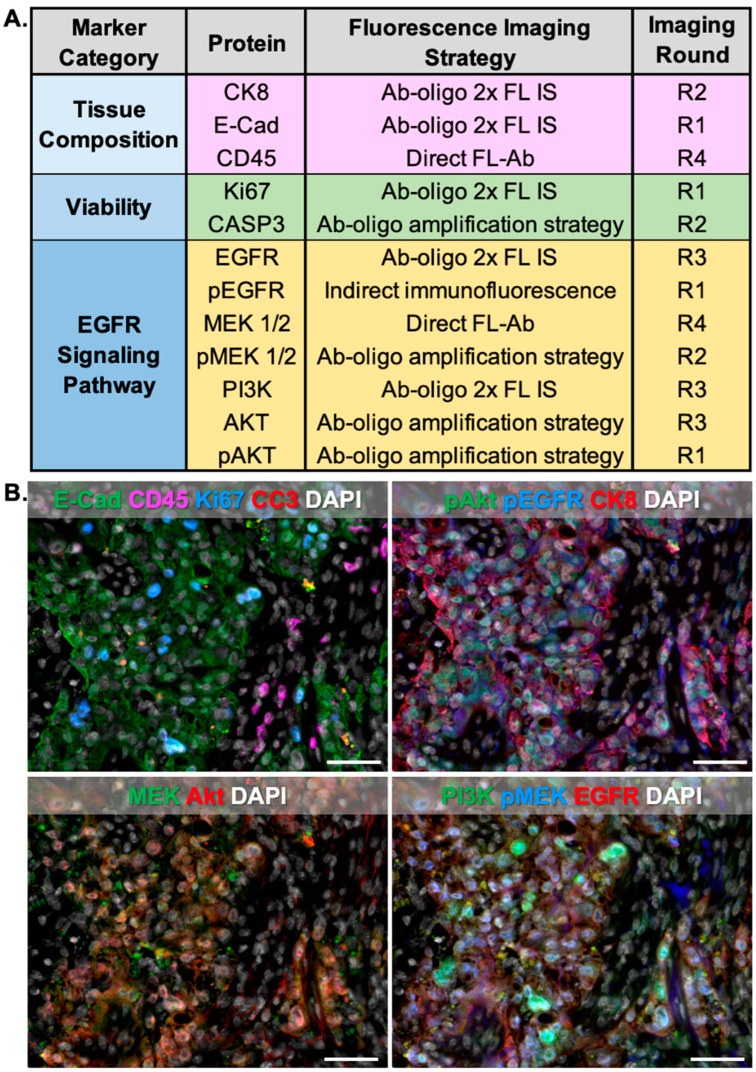
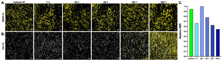
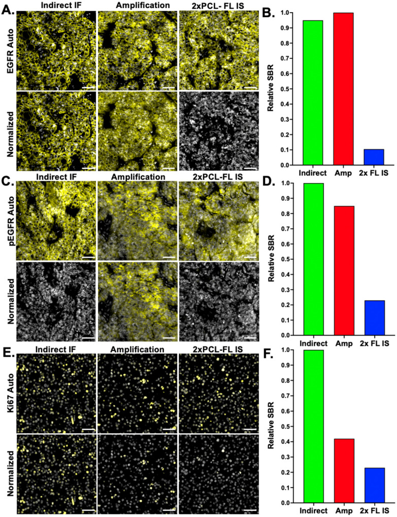
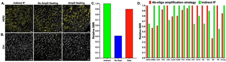
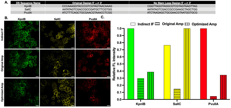
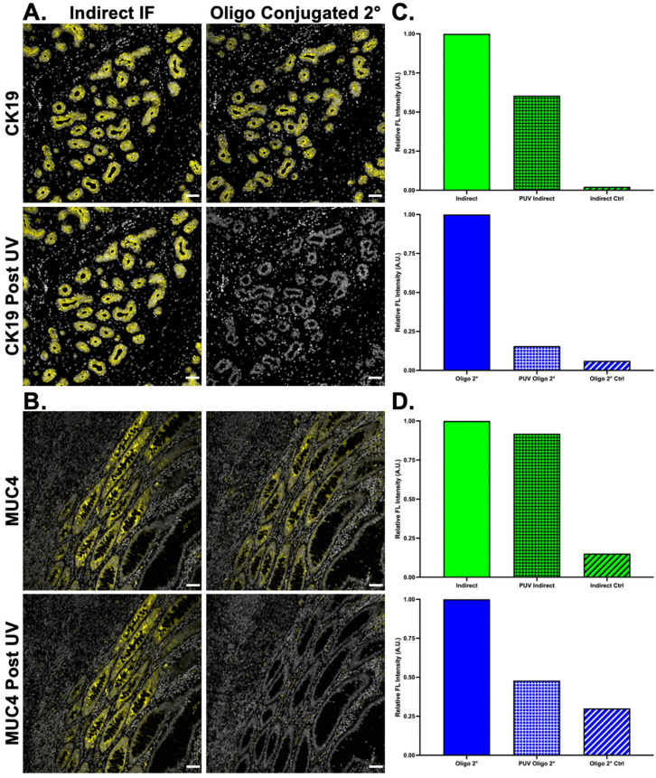
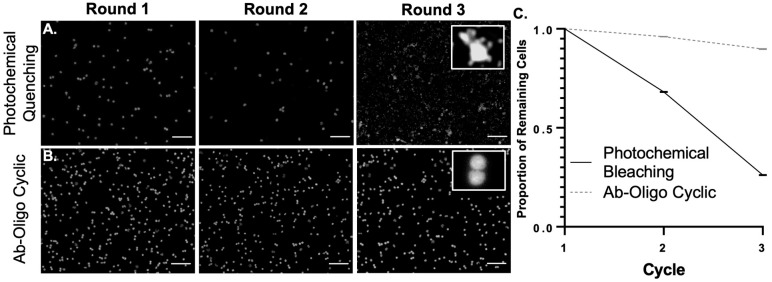
Advances in our understanding of the complex, multifaceted interactions between tumor epithelia, immune infiltrate, and tumor microenvironmental cells have been driven by highly multiplexed imaging technologies. These techniques are capable of labeling many more biomarkers than conventional immunostaining methods. However, multiplexed imaging techniques suffer from low detection sensitivity, cell loss-particularly in fragile samples-, and challenges with antibody labeling. Herein, we developed and optimized an oligonucleotide antibody barcoding strategy for cyclic immunofluorescence (cyCIF) that can be amplified to increase the detection efficiency of low-abundance antigens. Stained fluorescence signals can be readily removed using ultraviolet light treatment, preserving tissue and fragile cell sample integrity. We also extended the oligonucleotide barcoding strategy to secondary antibodies to enable the inclusion of difficult-to-label primary antibodies in a cyCIF panel. Using both the amplification oligonucleotides to label DNA barcoded antibodies and in situ hybridization of multiple fluorescently labeled oligonucleotides resulted in signal amplification and increased signal-to-background ratios. This procedure was optimized through the examination of staining parameters including staining oligonucleotide concentration, staining temperature, and oligonucleotide sequence design, resulting in a robust amplification technique. As a proof-of-concept, we demonstrate the flexibility of our cyCIF strategy by simultaneously imaging with the original oligonucleotide conjugated antibody (Ab-oligo) cyCIF strategy, the novel Ab-oligo cyCIF amplification strategy, as well as direct and indirect immunofluorescence to generate highly multiplexed images.
对肿瘤上皮细胞、免疫浸润细胞和肿瘤微环境细胞之间复杂多面相互作用的深入理解,得益于高度多重成像技术的发展。这些技术能够标记比传统免疫染色方法更多的生物标志物。然而,多重成像技术存在检测灵敏度低、细胞丢失(尤其是在脆弱样本中)以及抗体标记方面的挑战。在此,我们开发并优化了一种用于循环免疫荧光(cyCIF)的寡核苷酸抗体条形码策略,该策略可进行扩增以提高低丰度抗原的检测效率。使用紫外线处理可以轻松去除染色的荧光信号,同时保持组织和脆弱细胞样本的完整性。我们还将寡核苷酸条形码策略扩展到二抗,以便在cyCIF检测中纳入难以标记的一抗。使用扩增寡核苷酸标记DNA条形码抗体以及多种荧光标记寡核苷酸的原位杂交,实现了信号放大并提高了信噪比。通过检查染色参数,包括染色寡核苷酸浓度、染色温度和寡核苷酸序列设计,对该程序进行了优化,从而得到了一种可靠的扩增技术。作为概念验证,我们通过同时使用原始寡核苷酸偶联抗体(Ab - oligo)cyCIF策略、新型Ab - oligo cyCIF扩增策略以及直接和间接免疫荧光成像,展示了我们cyCIF策略的灵活性,以生成高度多重的图像。