Li Fan, Ma Lihua, Geng Yulei, Yan Xiaowei, Zhang Hengli, Tang Guangxian, Shang Qingli
Department of Ophthalmology, Shijiazhuang People's Hospital, Shijiazhuang 050000, Hebei, China.
Department of Ophthalmology, The Second Hospital of Hebei Medical University, Shijiazhuang 050000, Hebei, China.
J Ophthalmol. 2020 Nov 26;2020:8886398. doi: 10.1155/2020/8886398. eCollection 2020.
To evaluate the difference in macular choroidal thickness and volume between patients with pseudoexfoliative glaucoma (PXG), patients with pseudoexfoliative syndrome (PEX), and normal controls.
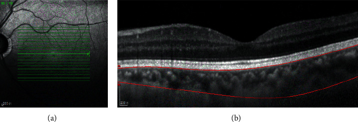
This case-control study included 49 PXG patients (group A), 33 PEX patients (group B), and 42 sex-, age-, and axial length-matched healthy volunteer eyes (group C). The macular choroidal thickness and volume of all subjects studied were measured by enhanced depth imaging optical coherence tomography.
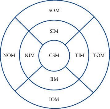
The average macular (AM) choroidal thickness was 170.79 ± 50.18 m, 184.65 ± 57.54 m, and 206.46 ± 48.90 m, and the average volume was 0.52 ± 0.15 m, 0.56 ± 0.17 m, and 0.63 ± 0.15 m in groups A, B, and C, respectively. The macular choroidal thickness, the volumes of various macular regions, and the average choroidal thickness and volume in group A were lower than those in group C (all < 0.05). There were no significant differences in the macular choroidal thickness, volumes of various macular regions, or average choroidal thickness or volume between group A and B (all > 0.05). The macular choroidal thickness and volume of the TIM and SOM in group B were lower than those in group C ( < 0.05). There was no association between the macular choroidal thickness of various macular regions and visual field mean defect (MD) in group A (all > 0.05).
The macular choroidal thickness in patients with PXG or PEX (TIM and SOM) is thinner than that in normal subjects. The macular choroidal thickness in patients with PXG is not significantly different from that in patients with PEX. The role of macular choroidal thickness changes in the glaucomatous damage of patients with PXG is still unclear.
评估假性剥脱性青光眼(PXG)患者、假性剥脱综合征(PEX)患者与正常对照组之间黄斑脉络膜厚度和体积的差异。
本病例对照研究纳入了49例PXG患者(A组)、33例PEX患者(B组)以及42只性别、年龄和眼轴长度匹配的健康志愿者眼睛(C组)。通过增强深度成像光学相干断层扫描测量所有研究对象的黄斑脉络膜厚度和体积。
A组、B组和C组的平均黄斑(AM)脉络膜厚度分别为170.79±50.18μm、184.65±57.54μm和206.46±48.90μm,平均体积分别为0.52±0.15μm、0.56±0.17μm和0.63±0.15μm。A组的黄斑脉络膜厚度、各个黄斑区域的体积以及平均脉络膜厚度和体积均低于C组(均P<0.05)。A组和B组在黄斑脉络膜厚度、各个黄斑区域的体积或平均脉络膜厚度和体积方面均无显著差异(均P>0.05)。B组颞下象限(TIM)和颞上象限(SOM)的黄斑脉络膜厚度和体积低于C组(P<0.05)。A组各个黄斑区域的黄斑脉络膜厚度与视野平均缺损(MD)之间无相关性(均P>0.05)。
PXG或PEX(TIM和SOM)患者的黄斑脉络膜厚度比正常受试者薄。PXG患者的黄斑脉络膜厚度与PEX患者无显著差异。黄斑脉络膜厚度变化在PXG患者青光眼性损害中的作用仍不清楚。