Nephrology and Transplantation, Department of Internal Medicine, Erasmus Medical Center, Rotterdam, Netherlands.
Microscopy CORE Lab, Maastricht Multimodal Molecular Imaging Institute, FHML Maastricht University, Maastricht, Netherlands.
Front Immunol. 2021 Apr 7;12:650522. doi: 10.3389/fimmu.2021.650522. eCollection 2021.
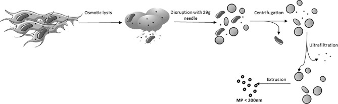
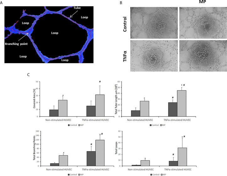
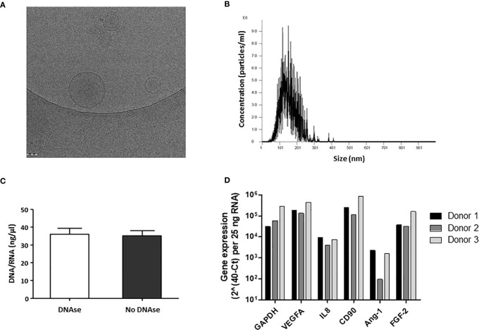
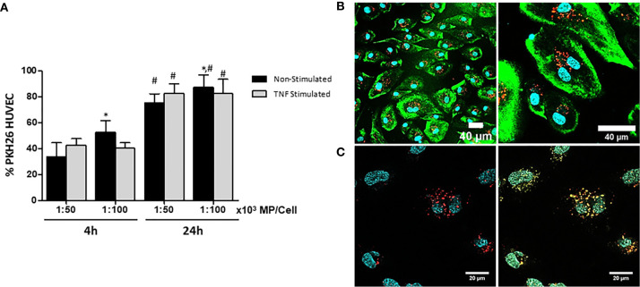
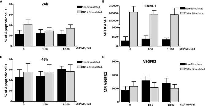
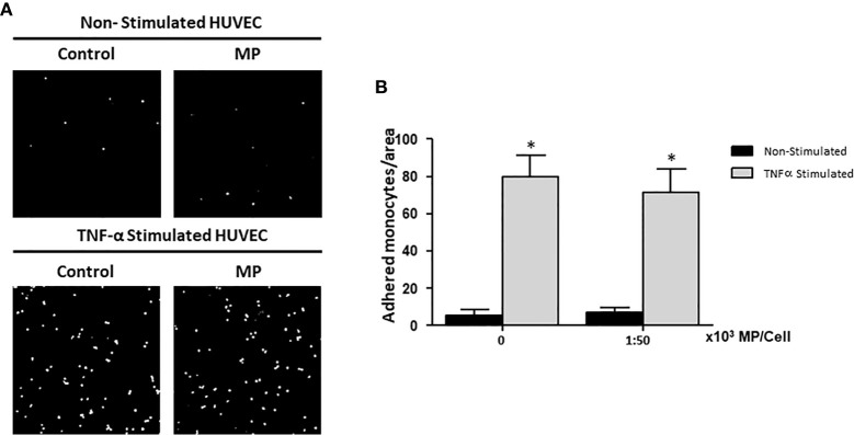
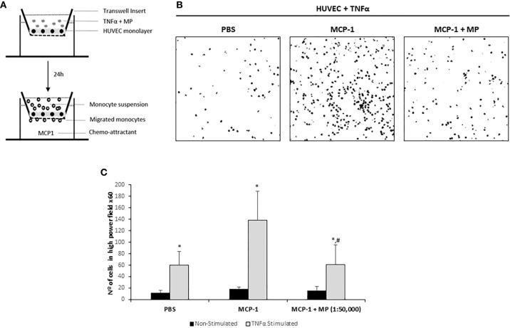
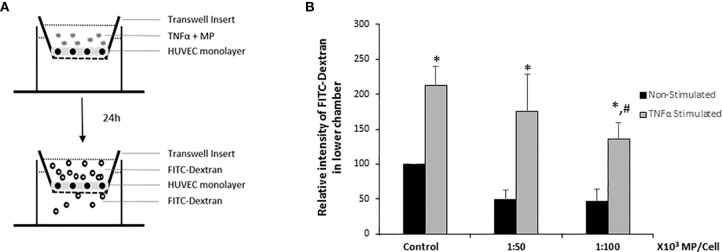
Proinflammatory stimuli lead to endothelial injury, which results in pathologies such as cardiovascular diseases, autoimmune diseases, and contributes to alloimmune responses after organ transplantation. Both mesenchymal stromal cells (MSC) and the extracellular vesicles (EV) released by them are widely studied as regenerative therapy for the endothelium. However, for therapeutic application, the manipulation of living MSC and large-scale production of EV are major challenges. Membrane particles (MP) generated from MSC may be an alternative to the use of whole MSC or EV. MP are nanovesicles artificially generated from the membranes of MSC and possess some of the therapeutic properties of MSC. In the present study we investigated whether MP conserve the beneficial MSC effects on endothelial cell repair processes under inflammatory conditions. MP were generated by hypotonic shock and extrusion of MSC membranes. The average size of MP was 120 nm, and they showed a spherical shape. The effects of two ratios of MP (50,000; 100,000 MP per target cell) on human umbilical vein endothelial cells (HUVEC) were tested in a model of inflammation induced by TNFα. Confocal microscopy and flow cytometry showed that within 24 hours >90% of HUVEC had taken up MP. Moreover, MP ended up in the lysosomes of the HUVEC. In a co-culture system of monocytes and TNFα activated HUVEC, MP did not affect monocyte adherence to HUVEC, but reduced the transmigration of monocytes across the endothelial layer from 138 ± 61 monocytes per microscopic field in TNFα activated HUVEC to 61 ± 45 monocytes. TNFα stimulation induced a 2-fold increase in the permeability of the HUVEC monolayer measured by the translocation of FITC-dextran to the lower compartment of a transwell system. At a dose of 1:100,000 MP significantly decreased endothelial permeability (1.5-fold) respect to TNFα Stimulated HUVEC. Finally, MP enhanced the angiogenic potential of HUVEC in an Matrigel assay by stimulating the formation of angiogenic structures, such as percentage of covered area, total tube length, total branching points, total loops. In conclusion, MP show regenerative effects on endothelial cells, opening a new avenue for treatment of vascular diseases where inflammatory processes damage the endothelium.
促炎刺激导致内皮损伤,从而导致心血管疾病、自身免疫性疾病等病理学发生,并导致器官移植后的同种免疫反应。间充质基质细胞(MSC)及其释放的细胞外囊泡(EV)都被广泛研究作为内皮的再生治疗方法。然而,对于治疗应用,对活 MSC 的操作和 EV 的大规模生产是主要挑战。源自 MSC 的膜颗粒(MP)可能是使用整个 MSC 或 EV 的替代方法。MP 是从 MSC 膜人工产生的纳米囊泡,具有 MSC 的一些治疗特性。在本研究中,我们研究了在炎症条件下,MP 是否保留了对内皮细胞修复过程的有益 MSC 作用。通过低渗冲击和 MSC 膜挤压来产生 MP。MP 的平均粒径为 120nm,呈球形。在 TNFα 诱导的炎症模型中,测试了两种 MP 比例(50000;每个靶细胞 100000 MP)对人脐静脉内皮细胞(HUVEC)的影响。共聚焦显微镜和流式细胞术显示,在 24 小时内,超过 90%的 HUVEC 摄取了 MP。此外,MP 最终进入 HUVEC 的溶酶体。在单核细胞和 TNFα 激活的 HUVEC 的共培养系统中,MP 不影响单核细胞与 HUVEC 的粘附,但减少了单核细胞穿过内皮层的迁移,从 TNFα 激活的 HUVEC 中每 100 个显微镜视野中的 138 ± 61 个单核细胞减少到 61 ± 45 个。TNFα 刺激导致 FITC-葡聚糖向 Transwell 系统下室的转移,使 HUVEC 单层的通透性增加 2 倍。在 1:100000 的剂量下,MP 与 TNFα 刺激的 HUVEC 相比,显著降低内皮通透性(1.5 倍)。最后,MP 通过刺激血管生成结构的形成,如覆盖面积百分比、总管长度、总分支点、总环数,在 Matrigel 测定中增强了 HUVEC 的血管生成潜力。总之,MP 对内皮细胞显示出再生作用,为治疗炎症过程损伤内皮的血管疾病开辟了新途径。