Joshi Bhagyashree S, de Beer Marit A, Giepmans Ben N G, Zuhorn Inge S
Department of Biomedical Engineering, University of Groningen, University Medical Center Groningen, 9713 AV Groningen, The Netherlands.
Department of Biomedical Sciences of Cells and Systems, University of Groningen, University Medical Center Groningen, 9713 AV Groningen, The Netherlands.
ACS Nano. 2020 Apr 28;14(4):4444-4455. doi: 10.1021/acsnano.9b10033. Epub 2020 Apr 16.
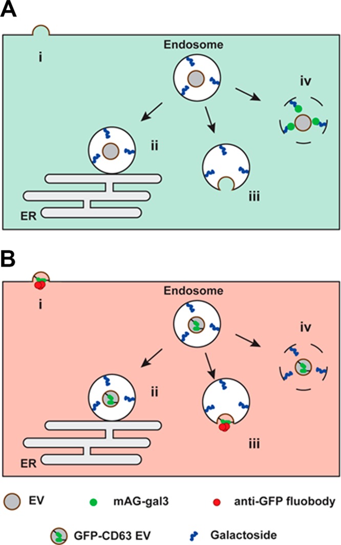
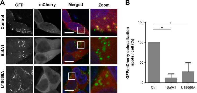
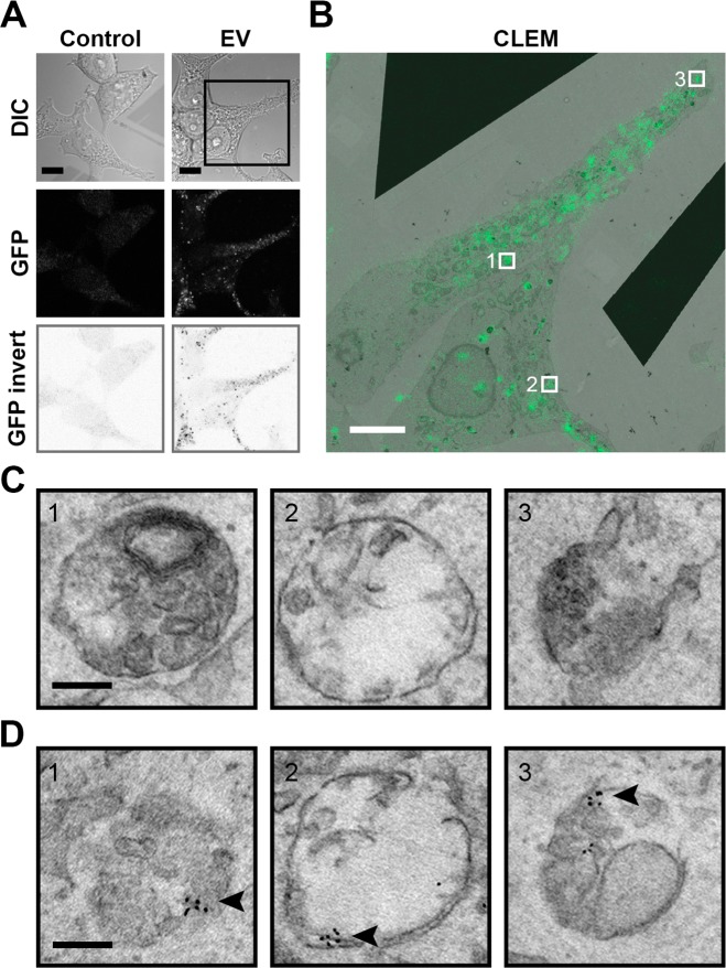
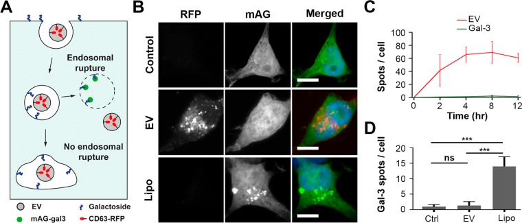
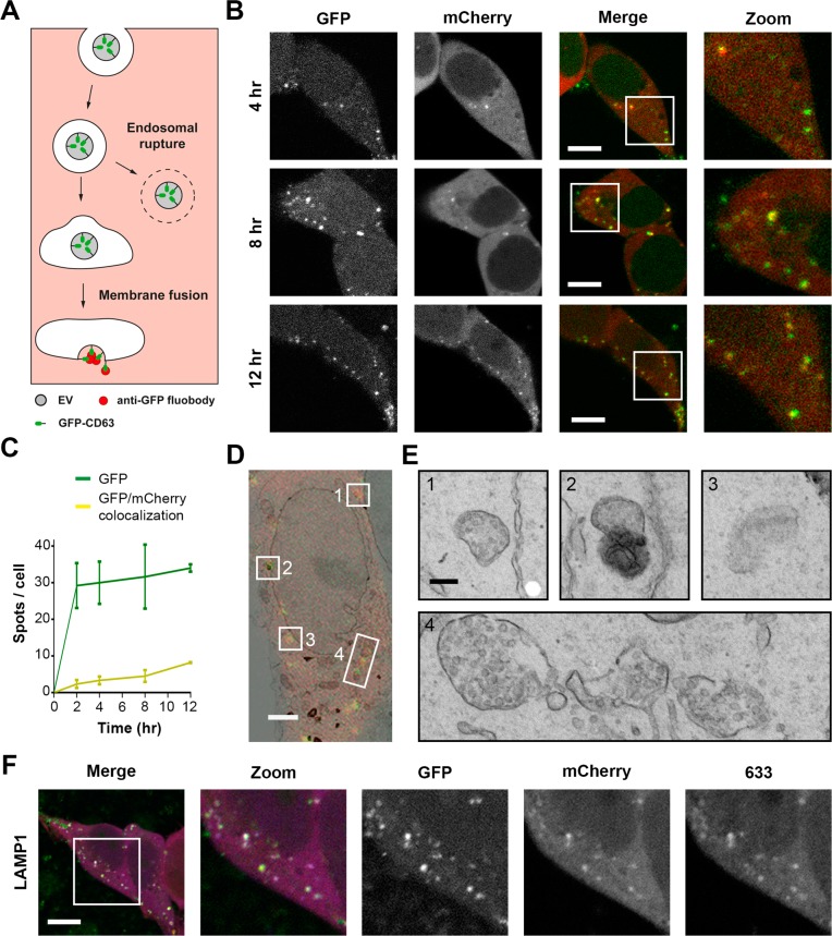
Extracellular vesicles (EVs), such as exosomes, can mediate long-distance communication between cells by delivering biomolecular cargo. It is speculated that EVs undergo back-fusion at multivesicular bodies (MVBs) in recipient cells to release their functional cargo. However, direct evidence is lacking. Tracing the cellular uptake of EVs with high resolution as well as acquiring direct evidence for the release of EV cargo is challenging mainly because of technical limitations. Here, we developed an analytical methodology, combining state-of-the-art molecular tools and correlative light and electron microscopy, to identify the intracellular site for EV cargo release. GFP was loaded inside EVs through the expression of GFP-CD63, a fusion of GFP to the cytosolic tail of CD63, in EV producer cells. In addition, we genetically engineered a cell line which expresses anti-GFP fluobody that specifically recognizes the EV cargo (GFP). Incubation of anti-GFP fluobody-expressing cells with GFP-CD63 EVs resulted in the formation of fluobody punctae, designating cytosolic exposure of GFP. Endosomal damage was not observed in EV acceptor cells. Ultrastructural analysis of the underlying structures at GFP/fluobody double-positive punctae demonstrated that EV cargo release occurs from endosomes/lysosomes. Finally, we show that neutralization of endosomal pH and cholesterol accumulation in endosomes leads to blockage of EV cargo exposure. In conclusion, we report that a fraction of internalized EVs fuse with the limiting membrane of endosomes/lysosomes in an acidification-dependent manner, which results in EV cargo exposure to the cell cytosol.
细胞外囊泡(EVs),如外泌体,可以通过传递生物分子货物来介导细胞间的长距离通讯。据推测,细胞外囊泡在受体细胞的多泡体(MVBs)处发生反向融合以释放其功能性货物。然而,缺乏直接证据。由于技术限制,以高分辨率追踪细胞外囊泡的细胞摄取以及获取细胞外囊泡货物释放的直接证据具有挑战性。在这里,我们开发了一种分析方法,结合了最先进的分子工具以及相关的光学和电子显微镜,以确定细胞外囊泡货物释放的细胞内位点。通过在细胞外囊泡产生细胞中表达GFP-CD63(一种GFP与CD63胞质尾部的融合蛋白),将GFP装载到细胞外囊泡内部。此外,我们对一个细胞系进行了基因工程改造,使其表达特异性识别细胞外囊泡货物(GFP)的抗GFP荧光抗体。将表达抗GFP荧光抗体的细胞与GFP-CD63细胞外囊泡孵育,导致荧光抗体斑点的形成,表明GFP在细胞质中暴露。在细胞外囊泡受体细胞中未观察到内体损伤。对GFP/荧光抗体双阳性斑点处的基础结构进行超微结构分析表明,细胞外囊泡货物从内体/溶酶体释放。最后,我们表明内体pH值的中和以及内体中胆固醇的积累会导致细胞外囊泡货物暴露的阻断。总之,我们报告内化的细胞外囊泡的一部分以酸化依赖的方式与内体/溶酶体的限制膜融合,这导致细胞外囊泡货物暴露于细胞质中。