Division of Molecular Biology, Ruđer Bošković Institute, Bijenička cesta 54, 10000 Zagreb, Croatia.
Division of Molecular Biology, Ruđer Bošković Institute, Bijenička cesta 54, 10000 Zagreb, Croatia.
Dev Cell. 2021 May 3;56(9):1253-1267.e10. doi: 10.1016/j.devcel.2021.04.005. Epub 2021 Apr 27.
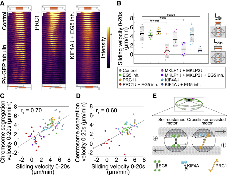
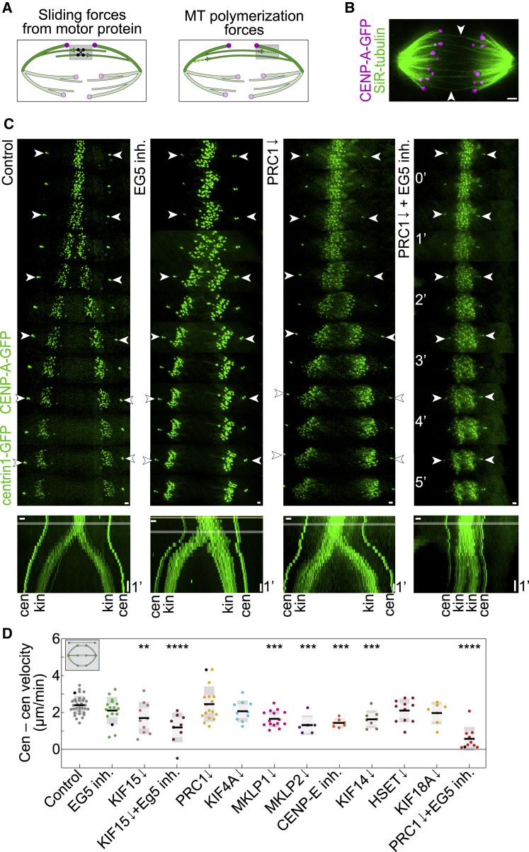
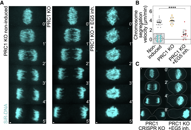
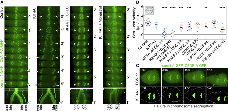
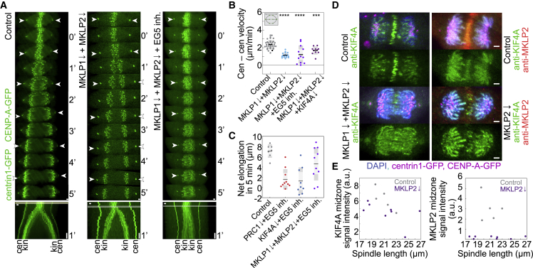
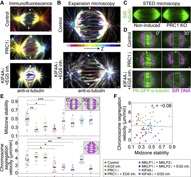
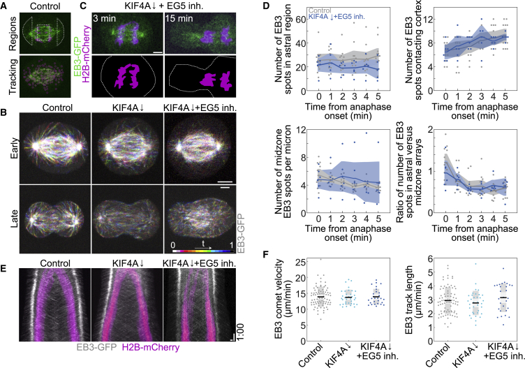
Proper chromosome segregation into two future daughter cells requires the mitotic spindle to elongate in anaphase. However, although some candidate proteins are implicated in this process, the molecular mechanism that drives spindle elongation in human cells is unknown. Using combined depletion and inactivation assays together with CRISPR technology to explore redundancy between multiple targets, we discovered that the force-generating mechanism of spindle elongation consists of EG5/kinesin-5 together with the PRC1-dependent motor KIF4A/kinesin-4, with contribution from kinesin-6 and kinesin-8. Disruption of EG5 and KIF4A leads to total failure of chromosome segregation due to blocked spindle elongation, despite poleward chromosome motion. Tubulin photoactivation, stimulated emission depletion (STED), and expansion microscopy show that perturbation of both proteins impairs midzone microtubule sliding without affecting microtubule stability. Thus, two mechanistically distinct sliding modules, one based on a self-sustained and the other on a crosslinker-assisted motor, power the mechanism that drives spindle elongation in human cells.
正确的染色体分离到两个未来的子细胞中需要有丝分裂纺锤体在后期拉长。然而,尽管一些候选蛋白与这个过程有关,但驱动人类细胞纺锤体伸长的分子机制尚不清楚。我们使用组合的耗尽和失活测定以及 CRISPR 技术来探索多个靶标之间的冗余性,发现纺锤体伸长的力产生机制由 EG5/驱动蛋白-5 与依赖 PRC1 的马达 KIF4A/驱动蛋白-4 组成,驱动蛋白-6 和驱动蛋白-8 也有贡献。尽管染色体向极运动,但 EG5 和 KIF4A 的破坏会导致纺锤体伸长受阻,导致染色体完全分离失败。微管光激活、受激发射损耗(STED)和扩展显微镜显示,两种蛋白的扰动会损害中间区微管的滑动,而不影响微管的稳定性。因此,两种机械上不同的滑动模块,一种基于自维持,另一种基于交联辅助马达,为驱动人类细胞纺锤体伸长的机制提供动力。