• 文献检索
  • 文档翻译
  • 深度研究
  • 学术资讯
  • Suppr Zotero 插件Zotero 插件
  • 邀请有礼
  • 套餐&价格
  • 历史记录
应用&插件
Suppr Zotero 插件Zotero 插件浏览器插件Mac 客户端Windows 客户端微信小程序
定价
高级版会员购买积分包购买API积分包
服务
文献检索文档翻译深度研究API 文档MCP 服务
关于我们
关于 Suppr公司介绍联系我们用户协议隐私条款
关注我们

Suppr 超能文献

核心技术专利:CN118964589B侵权必究
粤ICP备2023148730 号-1Suppr @ 2026

文献检索

告别复杂PubMed语法,用中文像聊天一样搜索,搜遍4000万医学文献。AI智能推荐,让科研检索更轻松。

立即免费搜索

文件翻译

保留排版,准确专业,支持PDF/Word/PPT等文件格式,支持 12+语言互译。

免费翻译文档

深度研究

AI帮你快速写综述,25分钟生成高质量综述,智能提取关键信息,辅助科研写作。

立即免费体验

周边视网膜光相干断层扫描血管造影在原发性开角型青光眼及周边正常眼的受影响和未受影响半球中的差异。

Circumpapillary optical coherence tomography angiography differences in perimetrically affected and unaffected hemispheres in primary open-angle glaucoma and the preperimetric fellow eye.

机构信息

Glaucoma Service, Dr Rajendra Prasad Centre for Ophthalmic Sciences, All India Institute of Medical Sciences, New Delhi, India.

出版信息

Indian J Ophthalmol. 2021 May;69(5):1120-1126. doi: 10.4103/ijo.IJO_1191_20.

DOI:10.4103/ijo.IJO_1191_20
PMID:33913845
原文链接:https://pmc.ncbi.nlm.nih.gov/articles/PMC8186637/
Abstract

PURPOSE

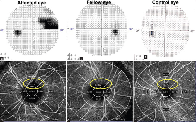
Evaluation of circumpapillary vessel density (VD) and perfusion density (PD) on optical coherence tomography angiography (OCTa) in mild-moderate glaucoma patients having unilateral visual field defects, with their fellow eyes and controls.

METHODS

Both eyes of 24 patients having a definitive nasal step or arcuate scotoma in one hemisphere of one eye only, and 24 controls, underwent OCTa.

RESULTS

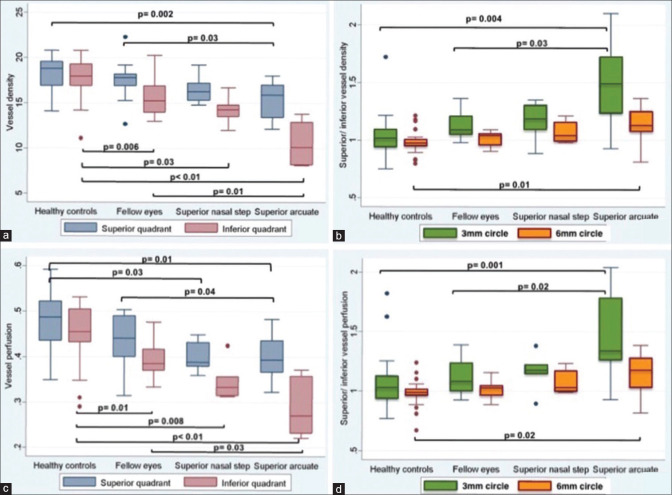
In eyes with a superior field defect, the superior/inferior quadrant ratios, (SQ/IQ) of 3 mm scan of VD and PD were significantly higher in eyes with a superior arcuate scotoma than fellow eyes (P = 0.03,0.02) as also controls, (P = 0.004,0.001). The mean percentage loss of inferior quadrant VD between control to fellow eyes, and superior nasal step eyes were similar, 20.19%/19.57% respectively, P = 0.85, while a loss in arcuate scotoma eyes was 38.81% (P = 0.001). The percentage decrease in inferior quadrant PD in fellow eyes was 14.70%, superior nasal step 23.39%, and an arcuate scotoma 34.74% (P = 0.02). Eyes with a superior nasal step had significantly lower VD and PD absolute values in the inferior quadrant OCTa in 3 mm and 6 mm circle scan only as compared to control eyes, VD, P = 0.03,0.04/PD, P = 0.008,0.02. Fellow eyes of superior field defects had significantly lower VD and PD absolute values in the inferior quadrant in 3 mm and 6 mm circle scan as compared to control eyes, VD, P = 0.006,0.04/PD, P = 0.01,0.03. Eyes with an isolated inferior field defect in only one eye, showed a significant decrease in both VD and PD in all quadrants as compared to fellow eyes and control eyes. A significant positive correlation was found between VD and RNFL thickness in peripapillary superior unaffected quadrants in eyes with superior field defects and inferior unaffected quadrants in inferior defects (P = 0.001 and 0.01).

CONCLUSION

There was a statistically significant increasing SQ/IQ ratio and percentage loss of vascular parameters from control to fellow eyes, those with a superior nasal step, and those with a superior arcuate scotoma. Inferior VFDs appeared to be associated with a more generalized circulatory loss. The asymmetry between hemispheres and between eyes could be used as a biomarker for early glaucomatous neuropathy.

摘要

目的

评估单侧视野缺损患者对侧眼和正常对照者的光学相干断层扫描血管造影(OCTa)的周边血管密度(VD)和灌注密度(PD)。

方法

对 24 例单侧视野缺损患者中只有一只眼存在明确的鼻侧阶梯或弓形暗点的双眼和 24 例对照者进行 OCTa 检查。

结果

在上方视野缺损的眼中,上方/下方象限比(SQ/IQ)在上方弓形暗点眼的 3mm 扫描中明显高于对侧眼(P=0.03,0.02),也高于对照组(P=0.004,0.001)。从对照组到对侧眼和上方鼻侧阶梯眼,下方象限 VD 的平均百分比损失相似,分别为 20.19%/19.57%,P=0.85,而在弓形暗点眼中的损失为 38.81%(P=0.001)。下方象限 PD 在对侧眼的下降百分比为 14.70%,上方鼻侧阶梯眼为 23.39%,弓形暗点眼为 34.74%(P=0.02)。只有上方鼻侧阶梯眼在 3mm 和 6mm 圆扫描中的下方象限 OCTa 中 VD 和 PD 的绝对值明显低于对照组,VD,P=0.03,0.04/PD,P=0.008,0.02。上方视野缺损的对侧眼在 3mm 和 6mm 圆扫描中的下方象限的 VD 和 PD 的绝对值明显低于对照组,VD,P=0.006,0.04/PD,P=0.01,0.03。只有一只眼存在孤立的下方视野缺损的眼,与对侧眼和对照组相比,所有象限的 VD 和 PD 均明显下降。在上方视野缺损的眼的上方未受影响的象限和下方视野缺损的眼的下方未受影响的象限中,发现 VD 和神经纤维层厚度之间存在显著的正相关(P=0.001 和 0.01)。

结论

从对照组到对侧眼、上方鼻侧阶梯眼和上方弓形暗点眼,血管参数的 SQ/IQ 比值和百分比损失均有统计学意义。下方 VFD 似乎与更广泛的循环损失有关。半球之间和眼之间的不对称性可以作为早期青光眼神经病变的生物标志物。

https://cdn.ncbi.nlm.nih.gov/pmc/blobs/7c8b/8186637/dbfd879b4fcc/IJO-69-1120-g002.jpg
https://cdn.ncbi.nlm.nih.gov/pmc/blobs/7c8b/8186637/4beb8600fbbb/IJO-69-1120-g001.jpg
https://cdn.ncbi.nlm.nih.gov/pmc/blobs/7c8b/8186637/dbfd879b4fcc/IJO-69-1120-g002.jpg
https://cdn.ncbi.nlm.nih.gov/pmc/blobs/7c8b/8186637/4beb8600fbbb/IJO-69-1120-g001.jpg
https://cdn.ncbi.nlm.nih.gov/pmc/blobs/7c8b/8186637/dbfd879b4fcc/IJO-69-1120-g002.jpg

相似文献

1
Circumpapillary optical coherence tomography angiography differences in perimetrically affected and unaffected hemispheres in primary open-angle glaucoma and the preperimetric fellow eye.周边视网膜光相干断层扫描血管造影在原发性开角型青光眼及周边正常眼的受影响和未受影响半球中的差异。
Indian J Ophthalmol. 2021 May;69(5):1120-1126. doi: 10.4103/ijo.IJO_1191_20.
2
Peripapillary and Macular Vascular Density in Patients With Preperimetric and Early Primary Open Angle Glaucoma.周边和黄斑血管密度在预旁中心视野和早期原发性开角型青光眼患者中的变化。
J Glaucoma. 2022 Sep 1;31(9):724-733. doi: 10.1097/IJG.0000000000002069. Epub 2022 Jun 21.
3
A Sectoral Analysis of Vessel Density Measurements in Perimetrically Intact Regions of Glaucomatous Eyes: An Optical Coherence Tomography Angiography Study.青光眼周边正常视野区血管密度的节段性分析:一项光学相干断层扫描血管造影研究。
J Glaucoma. 2018 Jun;27(6):525-531. doi: 10.1097/IJG.0000000000000950.
4
Optical coherence tomography angiography analysis of the fellow eye in unilateral pseudoexfoliation syndrome.单侧假性剥脱综合征对侧眼的光相干断层扫描血管造影分析。
BMC Ophthalmol. 2024 Sep 27;24(1):421. doi: 10.1186/s12886-024-03686-1.
5
Peripapillary Vessel Density and Retinal Nerve Fiber Layer Thickness in Patients with Unilateral Primary Angle Closure Glaucoma with Superior Hemifield Defect.单侧原发性闭角型青光眼伴上半视野缺损患者的视乳头周围血管密度和视网膜神经纤维层厚度
J Curr Glaucoma Pract. 2019 Jan-Apr;13(1):21-27. doi: 10.5005/jp-journals-10078-1247.
6
Comparison of microvascular parameters and diagnostic ability of optical coherence tomography angiography between eyes with primary angle closure glaucoma and primary open angle glaucoma.原发性闭角型青光眼与原发性开角型青光眼患者眼部的微血管参数及光学相干断层扫描血管造影诊断能力的比较
Photodiagnosis Photodyn Ther. 2022 Dec;40:103114. doi: 10.1016/j.pdpdt.2022.103114. Epub 2022 Sep 10.
7
Quantitative Analysis of Microvasculature in Macular and Peripapillary Regions in Early Primary Open-Angle Glaucoma.早期原发性开角型青光眼黄斑及视盘周围区域微血管定量分析。
Curr Eye Res. 2020 May;45(5):629-635. doi: 10.1080/02713683.2019.1676912. Epub 2019 Oct 14.
8
Projection-Resolved Optical Coherence Tomography Angiography of Macular Retinal Circulation in Glaucoma.青光眼黄斑视网膜循环的投影分辨光学相干断层扫描血管造影
Ophthalmology. 2017 Nov;124(11):1589-1599. doi: 10.1016/j.ophtha.2017.06.002. Epub 2017 Jul 1.
9
Asymmetry of Macular Vessel Density in Bilateral Early Open-angle Glaucoma With Unilateral Central 10-2 Visual Field Loss.双眼早期开角型青光眼伴单侧中央 10-2 视野损失的黄斑血管密度不对称性。
J Glaucoma. 2020 Oct;29(10):926-931. doi: 10.1097/IJG.0000000000001578.
10
Peripapillary choroidal thickness in both eyes of glaucoma patients with unilateral visual field loss.青光眼患者单侧视野缺损眼的周边脉络膜厚度。
Am J Ophthalmol. 2013 Dec;156(6):1277-1284.e1. doi: 10.1016/j.ajo.2013.07.011. Epub 2013 Sep 4.

引用本文的文献

1
Structural and functional differentiation between compressive and glaucomatous optic neuropathy.压迫性和青光眼性视神经病变的结构和功能分化。
Sci Rep. 2022 Apr 26;12(1):6795. doi: 10.1038/s41598-022-10269-x.
2
The Diagnostic Value of Pulsar Perimetry, Optical Coherence Tomography, and Optical Coherence Tomography Angiography in Pre-Perimetric and Perimetric Glaucoma.脉冲星视野检查、光学相干断层扫描及光学相干断层扫描血管造影在周边视野前及周边视野青光眼诊断中的价值
J Clin Med. 2021 Dec 13;10(24):5825. doi: 10.3390/jcm10245825.
3
Vessel Density in the Macular and Peripapillary Areas in Preperimetric Glaucoma to Various Stages of Primary Open-Angle Glaucoma in Taiwan.

本文引用的文献

1
Diagnostic Ability of Optical Coherence Tomography Angiography Macula Vessel Density for the Diagnosis of Glaucoma Using Difference Scan Sizes.利用不同大小的差分扫描来评估光学相干断层扫描血管造影黄斑血管密度诊断青光眼的能力。
J Glaucoma. 2020 Apr;29(4):245-251. doi: 10.1097/IJG.0000000000001447.
2
An overview of optical coherence tomography angiography and the posterior pole.光学相干断层扫描血管造影术与后极部概述。
Ther Adv Ophthalmol. 2019 Apr 3;11:2515841419840249. doi: 10.1177/2515841419840249. eCollection 2019 Jan-Dec.
3
Association of Macular and Circumpapillary Microvasculature with Visual Field Sensitivity in Advanced Glaucoma.
台湾青光眼前期黄斑区和视乳头周围区域血管密度与原发性开角型青光眼各阶段的关系
J Clin Med. 2021 Nov 23;10(23):5490. doi: 10.3390/jcm10235490.
4
Peripapillary optical coherence tomography angiography differences in perimetrically affected and unaffected hemispheres in primary open-angle glaucoma and the preperimetric fellow eye.原发性开角型青光眼视野受损和未受损半球以及视野前对侧眼的视乳头周围光学相干断层扫描血管造影差异
Indian J Ophthalmol. 2021 Oct;69(10):2894-2895. doi: 10.4103/ijo.IJO_1539_21.
高级青光眼患者黄斑和视盘周围微血管与视野敏感性的相关性研究。
Am J Ophthalmol. 2019 Aug;204:51-61. doi: 10.1016/j.ajo.2019.03.004. Epub 2019 Mar 14.
4
Measurement Floors and Dynamic Ranges of OCT and OCT Angiography in Glaucoma.青光眼 OCT 和 OCT 血管造影的测量下限和动态范围。
Ophthalmology. 2019 Jul;126(7):980-988. doi: 10.1016/j.ophtha.2019.03.003. Epub 2019 Mar 8.
5
Effect of Scan Size on Glaucoma Diagnostic Performance Using OCT Angiography En Face Images of the Radial Peripapillary Capillaries.基于 OCT 血管成像的放射状视神经周围毛细血管的扫描大小对青光眼诊断性能的影响。
J Glaucoma. 2019 May;28(5):465-472. doi: 10.1097/IJG.0000000000001216.
6
Diagnostic performance of optical coherence tomography angiography in glaucoma: a systematic review and meta-analysis.光学相干断层扫描血管造影术在青光眼诊断中的性能:系统评价和荟萃分析。
Br J Ophthalmol. 2019 Nov;103(11):1677-1684. doi: 10.1136/bjophthalmol-2018-313461. Epub 2019 Feb 6.
7
Diagnostic ability of macular ganglion cell asymmetry in Preperimetric Glaucoma.黄斑神经节细胞不对称性在青光眼前期视野检查中的诊断能力
BMC Ophthalmol. 2019 Jan 8;19(1):12. doi: 10.1186/s12886-018-1019-4.
8
Microvasculature of the Optic Nerve Head and Peripapillary Region in Patients With Primary Open-Angle Glaucoma.原发性开角型青光眼患者的视神经头和视盘周围区域的微血管。
J Glaucoma. 2019 Apr;28(4):281-288. doi: 10.1097/IJG.0000000000001165.
9
Diagnostic Performance of Macular Versus Peripapillary Vessel Parameters by Optical Coherence Tomography Angiography for Glaucoma.光学相干断层扫描血管造影术检测黄斑与视乳头周围血管参数对青光眼的诊断效能
Transl Vis Sci Technol. 2018 Dec 6;7(6):21. doi: 10.1167/tvst.7.6.21. eCollection 2018 Nov.
10
Peripapillary microvasculature in the retinal nerve fiber layer in glaucoma by optical coherence tomography angiography: focal structural and functional correlations and diagnostic performance.光学相干断层扫描血管造影术观察青光眼视网膜神经纤维层乳头周围微血管系统:局灶性结构与功能的相关性及诊断效能
Clin Ophthalmol. 2018 Nov 8;12:2285-2296. doi: 10.2147/OPTH.S179816. eCollection 2018.