Fachbereich Physik, Universität Hamburg and Center for Free-Electron Laser Science (CFEL), Luruper Chaussee 149, 22761 Hamburg, Germany.
Department of Chemistry, Universität Hamburg, Bundesstrasse 45, 20146 Hamburg, Germany.
Int J Mol Sci. 2021 Apr 1;22(7):3691. doi: 10.3390/ijms22073691.
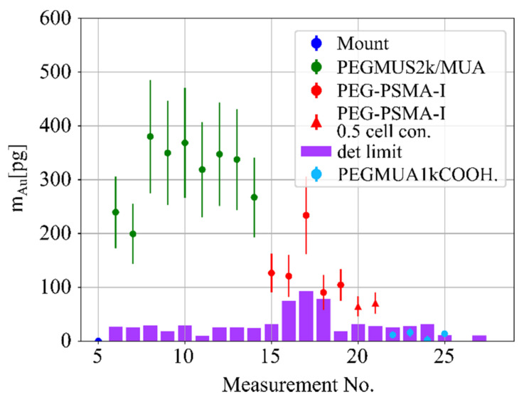
Quantitative cellular in vitro nanoparticle uptake measurements are possible with a large number of different techniques, however, all have their respective restrictions. Here, we demonstrate the application of synchrotron-based X-ray fluorescence imaging (XFI) on prostate tumor cells, which have internalized differently functionalized gold nanoparticles. Total nanoparticle uptake on the order of a few hundred picograms could be conveniently observed with microsamples consisting of only a few hundreds of cells. A comparison with mass spectroscopy quantification is provided, experimental results are both supported and sensitivity limits of this XFI approach extrapolated by Monte-Carlo simulations, yielding a minimum detectable nanoparticle mass of just 5 pg. This study demonstrates the high sensitivity level of XFI, allowing non-destructive uptake measurements with very small microsamples within just seconds of irradiation time.
定量细胞体外纳米颗粒摄取测量可以通过大量不同的技术来实现,然而,所有这些技术都有其各自的限制。在这里,我们展示了基于同步加速器的 X 射线荧光成像(XFI)在前列腺肿瘤细胞中的应用,这些细胞已经内化了不同功能化的金纳米颗粒。通过仅由几百个细胞组成的微样本,可以方便地观察到几百个皮克数量级的总纳米颗粒摄取。我们提供了与质谱定量的比较,实验结果得到了支持,并通过蒙特卡罗模拟推断出了这种 XFI 方法的灵敏度极限,得出了仅 5 皮克的最小可检测纳米颗粒质量。这项研究证明了 XFI 的高灵敏度水平,允许在仅几秒钟的辐照时间内,对非常小的微样本进行非破坏性摄取测量。