• 文献检索
  • 文档翻译
  • 深度研究
  • 学术资讯
  • Suppr Zotero 插件Zotero 插件
  • 邀请有礼
  • 套餐&价格
  • 历史记录
应用&插件
Suppr Zotero 插件Zotero 插件浏览器插件Mac 客户端Windows 客户端微信小程序
定价
高级版会员购买积分包购买API积分包
服务
文献检索文档翻译深度研究API 文档MCP 服务
关于我们
关于 Suppr公司介绍联系我们用户协议隐私条款
关注我们

Suppr 超能文献

核心技术专利:CN118964589B侵权必究
粤ICP备2023148730 号-1Suppr @ 2026

文献检索

告别复杂PubMed语法,用中文像聊天一样搜索,搜遍4000万医学文献。AI智能推荐,让科研检索更轻松。

立即免费搜索

文件翻译

保留排版,准确专业,支持PDF/Word/PPT等文件格式,支持 12+语言互译。

免费翻译文档

深度研究

AI帮你快速写综述,25分钟生成高质量综述,智能提取关键信息,辅助科研写作。

立即免费体验

基于对比增强光谱乳腺钼靶图像的放射组学特征降维方法预测乳腺癌

Radiomic Feature Reduction Approach to Predict Breast Cancer by Contrast-Enhanced Spectral Mammography Images.

作者信息

Massafra Raffaella, Bove Samantha, Lorusso Vito, Biafora Albino, Comes Maria Colomba, Didonna Vittorio, Diotaiuti Sergio, Fanizzi Annarita, Nardone Annalisa, Nolasco Angelo, Ressa Cosmo Maurizio, Tamborra Pasquale, Terenzio Antonella, La Forgia Daniele

机构信息

Struttura Semplice Dipartimentale di Fisica Sanitaria, I.R.C.C.S. Istituto Tumori "Giovanni Paolo II", Viale Orazio Flacco 65, 70124 Bari, Italy.

Dipartimento di Matematica, Università degli Studi di Bari, 70121 Bari, Italy.

出版信息

Diagnostics (Basel). 2021 Apr 10;11(4):684. doi: 10.3390/diagnostics11040684.

DOI:10.3390/diagnostics11040684
PMID:33920221
原文链接:https://pmc.ncbi.nlm.nih.gov/articles/PMC8070152/
Abstract

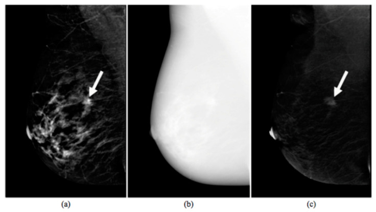
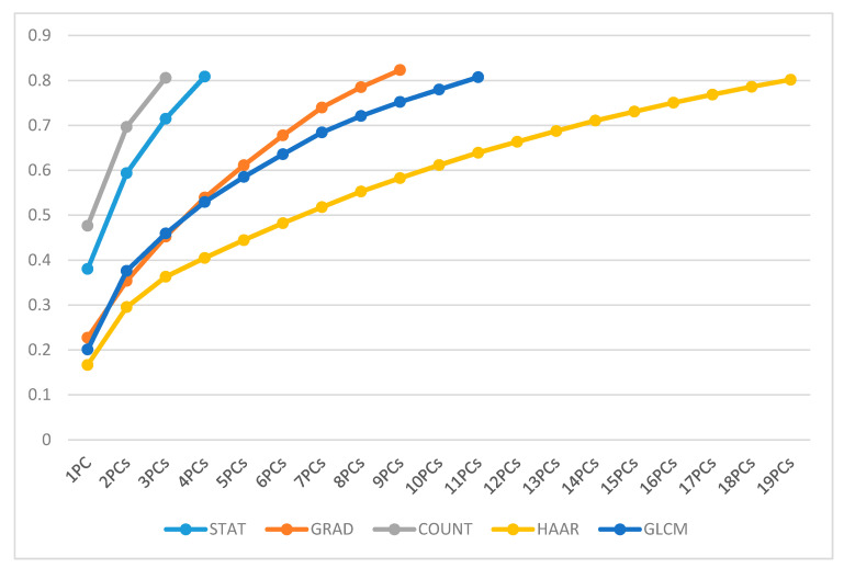
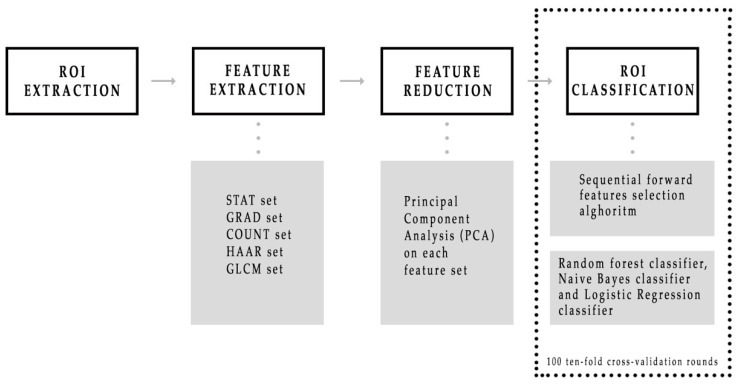
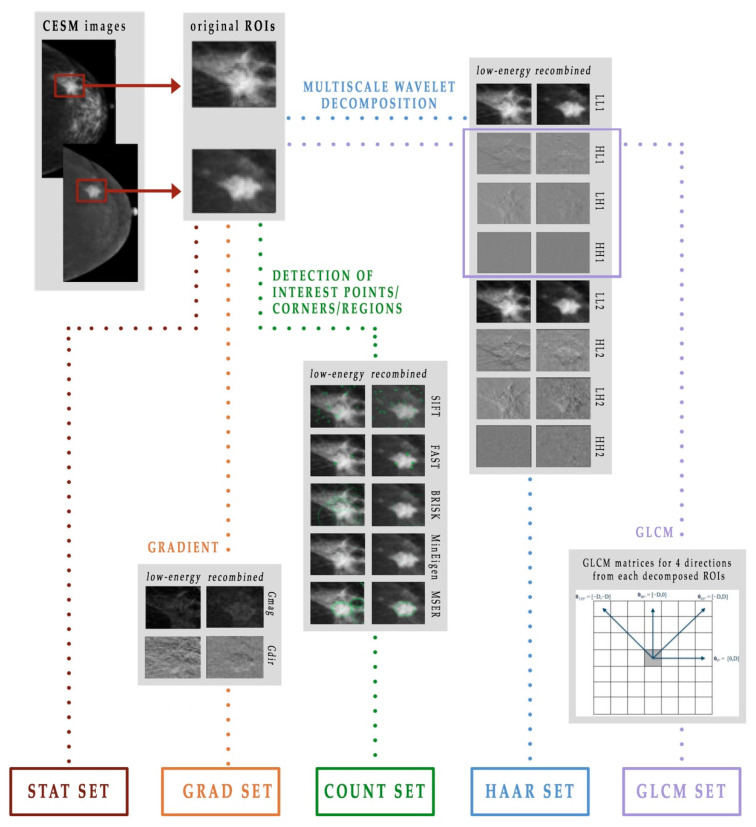
Contrast-enhanced spectral mammography (CESM) is an advanced instrument for breast care that is still operator dependent. The aim of this paper is the proposal of an automated system able to discriminate benign and malignant breast lesions based on radiomic analysis. We selected a set of 58 regions of interest (ROIs) extracted from 53 patients referred to Istituto Tumori "Giovanni Paolo II" of Bari (Italy) for the breast cancer screening phase between March 2017 and June 2018. We extracted 464 features of different kinds, such as points and corners of interest, textural and statistical features from both the original ROIs and the ones obtained by a Haar decomposition and a gradient image implementation. The features data had a large dimension that can affect the process and accuracy of cancer classification. Therefore, a classification scheme for dimension reduction was needed. Specifically, a principal component analysis (PCA) dimension reduction technique that includes the calculation of variance proportion for eigenvector selection was used. For the classification method, we trained three different classifiers, that is a random forest, a naïve Bayes and a logistic regression, on each sub-set of principal components (PC) selected by a sequential forward algorithm. Moreover, we focused on the starting features that contributed most to the calculation of the related PCs, which returned the best classification models. The method obtained with the aid of the random forest classifier resulted in the best prediction of benign/malignant ROIs with median values for sensitivity and specificity of 88.37% and 100%, respectively, by using only three PCs. The features that had shown the greatest contribution to the definition of the same were almost all extracted from the LE images. Our system could represent a valid support tool for radiologists for interpreting CESM images.

摘要

对比增强光谱乳腺摄影(CESM)是一种用于乳腺护理的先进仪器,但仍依赖操作人员。本文旨在提出一种基于放射组学分析能够区分乳腺良恶性病变的自动化系统。我们从2017年3月至2018年6月期间因乳腺癌筛查阶段转诊至意大利巴里“乔瓦尼·保罗二世”肿瘤研究所的53名患者中提取了一组58个感兴趣区域(ROI)。我们从原始ROI以及通过哈尔分解和梯度图像实现获得的ROI中提取了464种不同类型的特征,如兴趣点和角点、纹理和统计特征。特征数据维度很大,这可能会影响癌症分类的过程和准确性。因此,需要一种降维分类方案。具体而言,使用了一种主成分分析(PCA)降维技术,该技术包括计算用于特征向量选择的方差比例。对于分类方法,我们在通过顺序前向算法选择的每个主成分(PC)子集上训练了三种不同的分类器,即随机森林、朴素贝叶斯和逻辑回归。此外,我们关注对相关PC计算贡献最大的起始特征,这些特征返回了最佳分类模型。借助随机森林分类器获得的方法在仅使用三个PC的情况下,对良性/恶性ROI的预测效果最佳,灵敏度和特异性的中位数分别为88.37%和100%。对相同定义贡献最大的特征几乎都从LE图像中提取。我们的系统可以为放射科医生解释CESM图像提供有效的支持工具。

https://cdn.ncbi.nlm.nih.gov/pmc/blobs/2204/8070152/202327753533/diagnostics-11-00684-g005.jpg
https://cdn.ncbi.nlm.nih.gov/pmc/blobs/2204/8070152/d9ab5a9da0ef/diagnostics-11-00684-g001.jpg
https://cdn.ncbi.nlm.nih.gov/pmc/blobs/2204/8070152/fc3e1623a4d0/diagnostics-11-00684-g002.jpg
https://cdn.ncbi.nlm.nih.gov/pmc/blobs/2204/8070152/3dc0b9f9afe9/diagnostics-11-00684-g003.jpg
https://cdn.ncbi.nlm.nih.gov/pmc/blobs/2204/8070152/bf9b80bd9ef1/diagnostics-11-00684-g004.jpg
https://cdn.ncbi.nlm.nih.gov/pmc/blobs/2204/8070152/202327753533/diagnostics-11-00684-g005.jpg

相似文献

1
Radiomic Feature Reduction Approach to Predict Breast Cancer by Contrast-Enhanced Spectral Mammography Images.基于对比增强光谱乳腺钼靶图像的放射组学特征降维方法预测乳腺癌
Diagnostics (Basel). 2021 Apr 10;11(4):684. doi: 10.3390/diagnostics11040684.
2
Fully Automated Support System for Diagnosis of Breast Cancer in Contrast-Enhanced Spectral Mammography Images.用于对比增强光谱乳腺钼靶图像中乳腺癌诊断的全自动支持系统
J Clin Med. 2019 Jun 21;8(6):891. doi: 10.3390/jcm8060891.
3
Computer-aided diagnosis of contrast-enhanced spectral mammography: A feasibility study.计算机辅助诊断对比增强光谱乳腺摄影:一项可行性研究。
Eur J Radiol. 2018 Jan;98:207-213. doi: 10.1016/j.ejrad.2017.11.024. Epub 2017 Dec 5.
4
Classification of contrast-enhanced spectral mammography (CESM) images.对比增强光谱乳腺摄影(CESM)图像分类。
Int J Comput Assist Radiol Surg. 2019 Feb;14(2):249-257. doi: 10.1007/s11548-018-1876-6. Epub 2018 Oct 26.
5
A method for the automated classification of benign and malignant masses on digital breast tomosynthesis images using machine learning and radiomic features.一种利用机器学习和放射组学特征对数字乳腺断层合成图像上的良性和恶性肿块进行自动分类的方法。
Radiol Phys Technol. 2020 Mar;13(1):27-36. doi: 10.1007/s12194-019-00543-5. Epub 2019 Nov 4.
6
A machine learning approach on multiscale texture analysis for breast microcalcification diagnosis.基于多尺度纹理分析的机器学习方法在乳腺微钙化诊断中的应用。
BMC Bioinformatics. 2020 Mar 11;21(Suppl 2):91. doi: 10.1186/s12859-020-3358-4.
7
[Clinical value of suspicious calcification in the diagnosis and surgical treatment of breast lesions using contrast-enhanced spectral mammography].[利用对比增强光谱乳腺摄影术评估乳腺病变中可疑钙化在诊断及外科治疗中的临床价值]
Zhonghua Yi Xue Za Zhi. 2020 Jan 7;100(1):42-46. doi: 10.3760/cma.j.issn.0376-2491.2020.01.010.
8
Multiview multimodal network for breast cancer diagnosis in contrast-enhanced spectral mammography images.用于对比增强光谱乳腺钼靶图像中乳腺癌诊断的多视图多模态网络
Int J Comput Assist Radiol Surg. 2021 Jun;16(6):979-988. doi: 10.1007/s11548-021-02391-4. Epub 2021 May 8.
9
Prediction of Breast Cancer Histological Outcome by Radiomics and Artificial Intelligence Analysis in Contrast-Enhanced Mammography.在对比增强乳腺钼靶摄影中通过影像组学和人工智能分析预测乳腺癌组织学结果
Cancers (Basel). 2022 Apr 25;14(9):2132. doi: 10.3390/cancers14092132.
10
Differentiation of fat-poor angiomyolipoma from clear cell renal cell carcinoma in contrast-enhanced MDCT images using quantitative feature classification.基于定量特征分类的 MDCT 增强图像鉴别乏脂性血管平滑肌脂肪瘤与透明细胞肾细胞癌
Med Phys. 2017 Jul;44(7):3604-3614. doi: 10.1002/mp.12258. Epub 2017 Jun 9.

引用本文的文献

1
An updated overview of radiomics-based artificial intelligence (AI) methods in breast cancer screening and diagnosis.基于放射组学的人工智能(AI)方法在乳腺癌筛查和诊断中的最新综述。
Radiol Phys Technol. 2024 Dec;17(4):795-818. doi: 10.1007/s12194-024-00842-6. Epub 2024 Sep 16.
2
A multicentric study of radiomics and artificial intelligence analysis on contrast-enhanced mammography to identify different histotypes of breast cancer.多中心研究:基于对比增强乳腺摄影的影像组学和人工智能分析,以识别不同乳腺癌组织学分型。
Radiol Med. 2024 Jun;129(6):864-878. doi: 10.1007/s11547-024-01817-8. Epub 2024 May 17.
3
State-of-the-art for contrast-enhanced mammography.

本文引用的文献

1
Contrast-Enhanced Spectral Mammography-Based Radiomics Nomogram for the Prediction of Neoadjuvant Chemotherapy-Insensitive Breast Cancers.基于对比增强光谱乳腺摄影术的影像组学列线图预测新辅助化疗不敏感乳腺癌
Front Oncol. 2021 Feb 22;11:605230. doi: 10.3389/fonc.2021.605230. eCollection 2021.
2
Radiomic Analysis in Contrast-Enhanced Spectral Mammography for Predicting Breast Cancer Histological Outcome.对比增强光谱乳腺摄影术的放射组学分析用于预测乳腺癌组织学结果
Diagnostics (Basel). 2020 Sep 17;10(9):708. doi: 10.3390/diagnostics10090708.
3
Feasibility, Image Quality and Clinical Evaluation of Contrast-Enhanced Breast MRI Performed in a Supine Position Compared to the Standard Prone Position.
对比增强乳腺摄影的最新技术。
Br J Radiol. 2024 Mar 28;97(1156):695-704. doi: 10.1093/bjr/tqae017.
4
Breast MRI radiomics and machine learning-based predictions of response to neoadjuvant chemotherapy - How are they affected by variations in tumor delineation?基于乳腺MRI影像组学和机器学习的新辅助化疗反应预测——肿瘤勾画的变化如何影响它们?
Comput Struct Biotechnol J. 2023 Nov 19;23:52-63. doi: 10.1016/j.csbj.2023.11.016. eCollection 2024 Dec.
5
Radiomics and artificial intelligence analysis by T2-weighted imaging and dynamic contrast-enhanced magnetic resonance imaging to predict Breast Cancer Histological Outcome.基于 T2 加权成像和动态对比增强磁共振成像的放射组学和人工智能分析预测乳腺癌组织学结局。
Radiol Med. 2023 Nov;128(11):1347-1371. doi: 10.1007/s11547-023-01718-2. Epub 2023 Oct 6.
6
AI-Based Cancer Detection Model for Contrast-Enhanced Mammography.基于人工智能的对比增强乳腺X线摄影癌症检测模型
Bioengineering (Basel). 2023 Aug 17;10(8):974. doi: 10.3390/bioengineering10080974.
7
Identification and diagnosis of mammographic malignant architectural distortion using a deep learning based mask regional convolutional neural network.使用基于深度学习的掩码区域卷积神经网络对乳腺钼靶恶性结构扭曲进行识别与诊断。
Front Oncol. 2023 Mar 22;13:1119743. doi: 10.3389/fonc.2023.1119743. eCollection 2023.
8
Radiation-Free Microwave Technology for Breast Lesion Detection Using Supervised Machine Learning Model.基于监督机器学习模型的无辐射微波技术在乳腺病变检测中的应用。
Tomography. 2023 Jan 12;9(1):105-129. doi: 10.3390/tomography9010010.
9
Contrast-Enhanced Mammography (CEM) Capability to Distinguish Molecular Breast Cancer Subtypes.对比增强乳腺钼靶摄影(CEM)区分分子乳腺癌亚型的能力。
Biomedicines. 2022 Sep 24;10(10):2384. doi: 10.3390/biomedicines10102384.
10
A machine learning ensemble approach for 5- and 10-year breast cancer invasive disease event classification.机器学习集成方法用于 5 年和 10 年乳腺癌浸润性疾病事件分类。
PLoS One. 2022 Sep 19;17(9):e0274691. doi: 10.1371/journal.pone.0274691. eCollection 2022.
仰卧位对比增强乳腺MRI与标准俯卧位对比增强乳腺MRI的可行性、图像质量及临床评估
Cancers (Basel). 2020 Aug 21;12(9):2364. doi: 10.3390/cancers12092364.
4
A machine learning approach on multiscale texture analysis for breast microcalcification diagnosis.基于多尺度纹理分析的机器学习方法在乳腺微钙化诊断中的应用。
BMC Bioinformatics. 2020 Mar 11;21(Suppl 2):91. doi: 10.1186/s12859-020-3358-4.
5
Microcalcification detection in full-field digital mammograms: A fully automated computer-aided system.全数字化乳腺钼靶片中微钙化的检测:一种全自动计算机辅助系统。
Phys Med. 2019 Aug;64:1-9. doi: 10.1016/j.ejmp.2019.05.022. Epub 2019 Jun 14.
6
Logistic Regression for Machine Learning in Process Tomography.过程层析成像中用于机器学习的逻辑回归
Sensors (Basel). 2019 Aug 2;19(15):3400. doi: 10.3390/s19153400.
7
Diagnostic performance of contrast-enhanced dual-energy spectral mammography (CESM): a retrospective study involving 644 breast lesions.对比增强双能光谱乳腺摄影(CESM)的诊断性能:一项涉及 644 个乳腺病变的回顾性研究。
Radiol Med. 2019 Oct;124(10):1006-1017. doi: 10.1007/s11547-019-01056-2. Epub 2019 Jun 27.
8
Fully Automated Support System for Diagnosis of Breast Cancer in Contrast-Enhanced Spectral Mammography Images.用于对比增强光谱乳腺钼靶图像中乳腺癌诊断的全自动支持系统
J Clin Med. 2019 Jun 21;8(6):891. doi: 10.3390/jcm8060891.
9
Breast MRI background parenchymal enhancement as an imaging bridge to molecular cancer sub-type.乳腺 MRI 背景实质增强作为连接分子肿瘤亚型的影像学桥梁。
Eur J Radiol. 2019 Apr;113:148-152. doi: 10.1016/j.ejrad.2019.02.018. Epub 2019 Feb 15.
10
Detection of Breast Cancer with Mammography: Effect of an Artificial Intelligence Support System.乳腺 X 线摄影术检测乳腺癌:人工智能支持系统的效果。
Radiology. 2019 Feb;290(2):305-314. doi: 10.1148/radiol.2018181371. Epub 2018 Nov 20.