• 文献检索
  • 文档翻译
  • 深度研究
  • 学术资讯
  • Suppr Zotero 插件Zotero 插件
  • 邀请有礼
  • 套餐&价格
  • 历史记录
应用&插件
Suppr Zotero 插件Zotero 插件浏览器插件Mac 客户端Windows 客户端微信小程序
定价
高级版会员购买积分包购买API积分包
服务
文献检索文档翻译深度研究API 文档MCP 服务
关于我们
关于 Suppr公司介绍联系我们用户协议隐私条款
关注我们

Suppr 超能文献

核心技术专利:CN118964589B侵权必究
粤ICP备2023148730 号-1Suppr @ 2026

文献检索

告别复杂PubMed语法,用中文像聊天一样搜索,搜遍4000万医学文献。AI智能推荐,让科研检索更轻松。

立即免费搜索

文件翻译

保留排版,准确专业,支持PDF/Word/PPT等文件格式,支持 12+语言互译。

免费翻译文档

深度研究

AI帮你快速写综述,25分钟生成高质量综述,智能提取关键信息,辅助科研写作。

立即免费体验

使用标准镶嵌语言模型在磁共振成像中基于卷积神经网络的颅骨分割技术的开发。

Development of a Convolutional Neural Network Based Skull Segmentation in MRI Using Standard Tesselation Language Models.

作者信息

Dalvit Carvalho da Silva Rodrigo, Jenkyn Thomas Richard, Carranza Victor Alexander

机构信息

Craniofacial Injury and Concussion Research Laboratory, Western University, London, ON N6A 3K7, Canada.

Faculty of Engineering, School of Biomedical Engineering, Western University, London, ON N6A 3K7, Canada.

出版信息

J Pers Med. 2021 Apr 16;11(4):310. doi: 10.3390/jpm11040310.

DOI:10.3390/jpm11040310
PMID:33923480
原文链接:https://pmc.ncbi.nlm.nih.gov/articles/PMC8074044/
Abstract

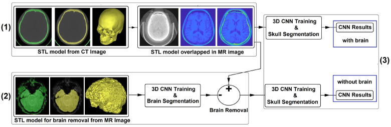
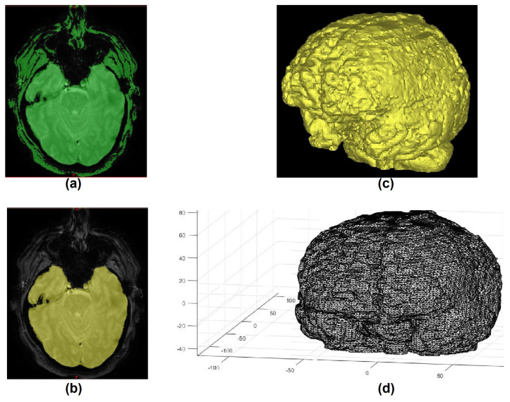
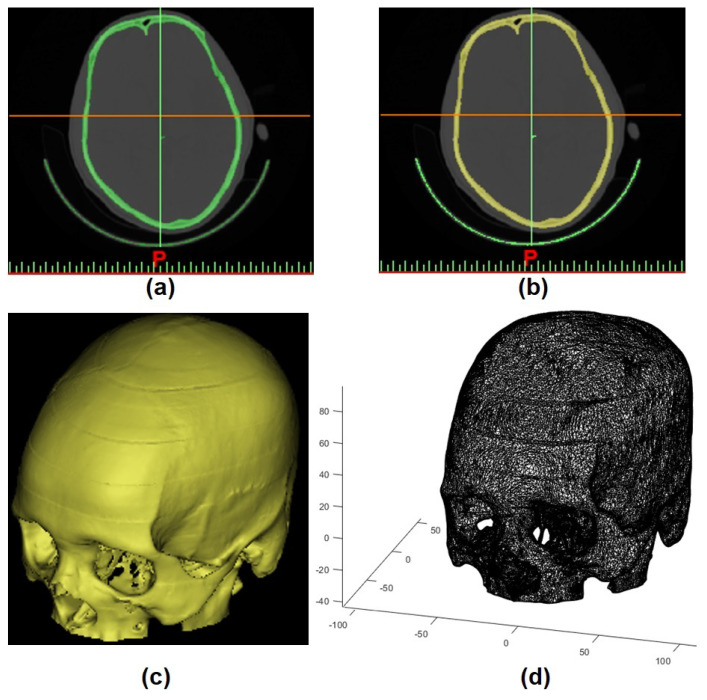
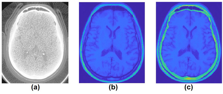
Segmentation is crucial in medical imaging analysis to help extract regions of interest (ROI) from different imaging modalities. The aim of this study is to develop and train a 3D convolutional neural network (CNN) for skull segmentation in magnetic resonance imaging (MRI). 58 gold standard volumetric labels were created from computed tomography (CT) scans in standard tessellation language (STL) models. These STL models were converted into matrices and overlapped on the 58 corresponding MR images to create the MRI gold standards labels. The CNN was trained with these 58 MR images and a mean ± standard deviation (SD) Dice similarity coefficient (DSC) of 0.7300 ± 0.04 was achieved. A further investigation was carried out where the brain region was removed from the image with the help of a 3D CNN and manual corrections by using only MR images. This new dataset, without the brain, was presented to the previous CNN which reached a new mean ± SD DSC of 0.7826 ± 0.03. This paper aims to provide a framework for segmenting the skull using CNN and STL models, as the 3D CNN was able to segment the skull with a certain precision.

摘要

在医学影像分析中,分割对于从不同成像模态中提取感兴趣区域(ROI)至关重要。本研究的目的是开发并训练一个用于磁共振成像(MRI)中颅骨分割的三维卷积神经网络(CNN)。从计算机断层扫描(CT)扫描的标准镶嵌语言(STL)模型中创建了58个金标准体积标签。这些STL模型被转换为矩阵,并与58幅相应的MR图像重叠,以创建MRI金标准标签。使用这58幅MR图像对CNN进行训练,平均±标准差(SD)的骰子相似系数(DSC)达到了0.7300±0.04。进一步的研究是在仅使用MR图像的情况下,借助三维CNN和手动校正从图像中去除脑区。这个没有脑区的新数据集被提供给之前的CNN,其新的平均±标准差DSC达到了0.7826±0.03。本文旨在提供一个使用CNN和STL模型分割颅骨的框架,因为三维CNN能够以一定精度分割颅骨。

https://cdn.ncbi.nlm.nih.gov/pmc/blobs/6927/8074044/bbf9c284b338/jpm-11-00310-g004.jpg
https://cdn.ncbi.nlm.nih.gov/pmc/blobs/6927/8074044/f10801bb7a31/jpm-11-00310-g001.jpg
https://cdn.ncbi.nlm.nih.gov/pmc/blobs/6927/8074044/f756e03f5579/jpm-11-00310-g002.jpg
https://cdn.ncbi.nlm.nih.gov/pmc/blobs/6927/8074044/24b7253283dd/jpm-11-00310-g003.jpg
https://cdn.ncbi.nlm.nih.gov/pmc/blobs/6927/8074044/bbf9c284b338/jpm-11-00310-g004.jpg

相似文献

1
Development of a Convolutional Neural Network Based Skull Segmentation in MRI Using Standard Tesselation Language Models.使用标准镶嵌语言模型在磁共振成像中基于卷积神经网络的颅骨分割技术的开发。
J Pers Med. 2021 Apr 16;11(4):310. doi: 10.3390/jpm11040310.
2
CT image segmentation of bone for medical additive manufacturing using a convolutional neural network.基于卷积神经网络的医学增材制造中骨骼的 CT 图像分割。
Comput Biol Med. 2018 Dec 1;103:130-139. doi: 10.1016/j.compbiomed.2018.10.012. Epub 2018 Oct 16.
3
Deep learning approaches using 2D and 3D convolutional neural networks for generating male pelvic synthetic computed tomography from magnetic resonance imaging.使用 2D 和 3D 卷积神经网络从磁共振成像生成男性骨盆合成 CT 的深度学习方法。
Med Phys. 2019 Sep;46(9):3788-3798. doi: 10.1002/mp.13672. Epub 2019 Jul 26.
4
Convolutional neural network-based approach for segmentation of left ventricle myocardial scar from 3D late gadolinium enhancement MR images.基于卷积神经网络的方法用于从 3D 晚期钆增强磁共振图像中分割左心室心肌瘢痕。
Med Phys. 2019 Apr;46(4):1740-1751. doi: 10.1002/mp.13436. Epub 2019 Feb 28.
5
Segmentation of dental cone-beam CT scans affected by metal artifacts using a mixed-scale dense convolutional neural network.使用混合尺度密集卷积神经网络对受金属伪影影响的口腔锥形束 CT 扫描进行分割。
Med Phys. 2019 Nov;46(11):5027-5035. doi: 10.1002/mp.13793. Epub 2019 Sep 13.
6
Cross-modality deep learning: Contouring of MRI data from annotated CT data only.跨模态深度学习:仅从标注的CT数据对MRI数据进行轮廓提取。
Med Phys. 2021 Apr;48(4):1673-1684. doi: 10.1002/mp.14619. Epub 2020 Dec 13.
7
Automated segmentation of the human supraclavicular fat depot via deep neural network in water-fat separated magnetic resonance images.通过深度神经网络在水脂分离磁共振图像中对人体锁骨上脂肪库进行自动分割。
Quant Imaging Med Surg. 2023 Jul 1;13(7):4699-4715. doi: 10.21037/qims-22-304. Epub 2023 Mar 14.
8
Automatic segmentation of high-risk clinical target volume for tandem-and-ovoids brachytherapy patients using an asymmetric dual-path convolutional neural network.使用非对称双通道卷积神经网络对 tandem-and-ovoids 近距离治疗患者的高危临床靶区进行自动分割。
Med Phys. 2022 Mar;49(3):1712-1722. doi: 10.1002/mp.15490. Epub 2022 Feb 4.
9
Two-stage deep learning model for fully automated pancreas segmentation on computed tomography: Comparison with intra-reader and inter-reader reliability at full and reduced radiation dose on an external dataset.基于 CT 的全自动胰腺分割的两阶段深度学习模型:在外部数据集上比较全剂量和低剂量下的同读者和异读者可靠性。
Med Phys. 2021 May;48(5):2468-2481. doi: 10.1002/mp.14782. Epub 2021 Mar 16.
10
Volumetric Pancreas Segmentation on Computed Tomography: Accuracy and Efficiency of a Convolutional Neural Network Versus Manual Segmentation in 3D Slicer in the Context of Interreader Variability of Expert Radiologists.体素胰腺 CT 分割:卷积神经网络与专家放射科医生 3D Slicer 中手动分割在读者间变异性方面的准确性和效率比较。
J Comput Assist Tomogr. 2022;46(6):841-847. doi: 10.1097/RCT.0000000000001374. Epub 2022 Sep 1.

引用本文的文献

1
NEC-NET : Segmentation and Feature Extraction Network for the Neurocranium in Early Childhood.NEC-NET:幼儿期颅盖骨分割与特征提取网络
Proc SPIE Int Soc Opt Eng. 2022 Nov;12567. doi: 10.1117/12.2670281. Epub 2023 Mar 6.
2
Deep Learning-Based Automated Magnetic Resonance Image Segmentation of the Lumbar Structure and Its Adjacent Structures at the L4/5 Level.基于深度学习的L4/5水平腰椎结构及其相邻结构的磁共振图像自动分割
Bioengineering (Basel). 2023 Aug 15;10(8):963. doi: 10.3390/bioengineering10080963.
3
Use of Advanced Artificial Intelligence in Forensic Medicine, Forensic Anthropology and Clinical Anatomy.

本文引用的文献

1
Generalised Dice Overlap as a Deep Learning Loss Function for Highly Unbalanced Segmentations.广义骰子重叠作为高度不平衡分割的深度学习损失函数
Deep Learn Med Image Anal Multimodal Learn Clin Decis Support (2017). 2017;2017:240-248. doi: 10.1007/978-3-319-67558-9_28. Epub 2017 Sep 9.
2
Convolutional Neural Networks and Geometric Moments to Identify the Bilateral Symmetric Midplane in Facial Skeletons from CT Scans.卷积神经网络与几何矩用于从CT扫描中识别面部骨骼的双侧对称中平面
Biology (Basel). 2021 Mar 2;10(3):182. doi: 10.3390/biology10030182.
3
Assessment of Compatibility between Various Intraoral Scanners and 3D Printers through an Accuracy Analysis of 3D Printed Models.
先进人工智能在法医学、法医人类学和临床解剖学中的应用。
Healthcare (Basel). 2021 Nov 12;9(11):1545. doi: 10.3390/healthcare9111545.
通过3D打印模型的精度分析评估各种口腔内扫描仪与3D打印机之间的兼容性
Materials (Basel). 2020 Oct 4;13(19):4419. doi: 10.3390/ma13194419.
4
UNet++: A Nested U-Net Architecture for Medical Image Segmentation.U-Net++:一种用于医学图像分割的嵌套U-Net架构。
Deep Learn Med Image Anal Multimodal Learn Clin Decis Support (2018). 2018 Sep;11045:3-11. doi: 10.1007/978-3-030-00889-5_1. Epub 2018 Sep 20.
5
Novel Digital Technique to Quantify the Area and Volume of Cement Remaining and Enamel Removed after Fixed Multibracket Appliance Therapy Debonding: An In Vitro Study.一种用于量化固定多托槽矫治器拆除后剩余黏固剂面积和去除牙釉质体积的新型数字技术:一项体外研究。
J Clin Med. 2020 Apr 12;9(4):1098. doi: 10.3390/jcm9041098.
6
Deep Learning for Cardiac Image Segmentation: A Review.用于心脏图像分割的深度学习:综述
Front Cardiovasc Med. 2020 Mar 5;7:25. doi: 10.3389/fcvm.2020.00025. eCollection 2020.
7
Reducing the Hausdorff Distance in Medical Image Segmentation With Convolutional Neural Networks.基于卷积神经网络的医学图像分割中的 Hausdorff 距离减少。
IEEE Trans Med Imaging. 2020 Feb;39(2):499-513. doi: 10.1109/TMI.2019.2930068. Epub 2019 Jul 19.
8
Image Thresholding Improves 3-Dimensional Convolutional Neural Network Diagnosis of Different Acute Brain Hemorrhages on Computed Tomography Scans.图像阈值处理提高 CT 扫描下不同类型急性脑出血的三维卷积神经网络诊断效果。
Sensors (Basel). 2019 May 10;19(9):2167. doi: 10.3390/s19092167.
9
A U-Net Deep Learning Framework for High Performance Vessel Segmentation in Patients With Cerebrovascular Disease.一种用于脑血管疾病患者高性能血管分割的U-Net深度学习框架。
Front Neurosci. 2019 Feb 28;13:97. doi: 10.3389/fnins.2019.00097. eCollection 2019.
10
Automated segmentation of knee bone and cartilage combining statistical shape knowledge and convolutional neural networks: Data from the Osteoarthritis Initiative.基于统计形状知识和卷积神经网络的膝关节骨和软骨自动分割:来自 Osteoarthritis Initiative 的数据。
Med Image Anal. 2019 Feb;52:109-118. doi: 10.1016/j.media.2018.11.009. Epub 2018 Nov 17.