Department of Cardiology, Cardiology I-Laboratory of Molecular Cardiology, University Medical Center of the Johannes Gutenberg-University, Building 605, Langenbeckstr. 1, 55131, Mainz, Germany.
Department of Ophthalmology, University Medical Center of the Johannes Gutenberg University, Mainz, Germany.
Basic Res Cardiol. 2021 Apr 30;116(1):31. doi: 10.1007/s00395-021-00869-5.
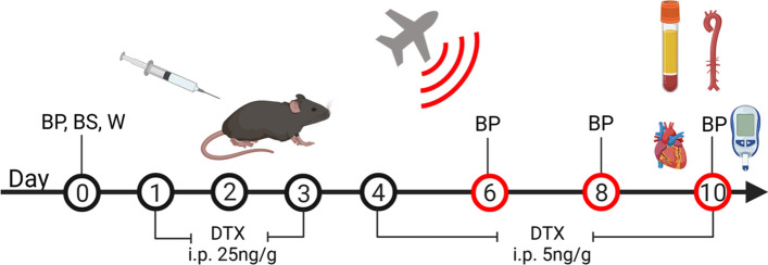
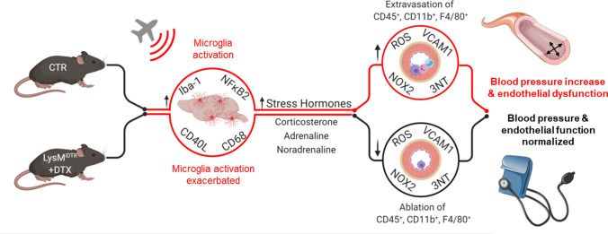
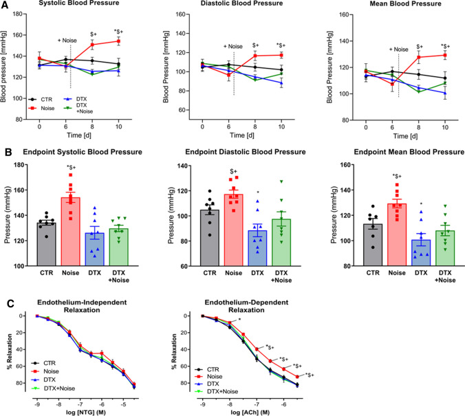
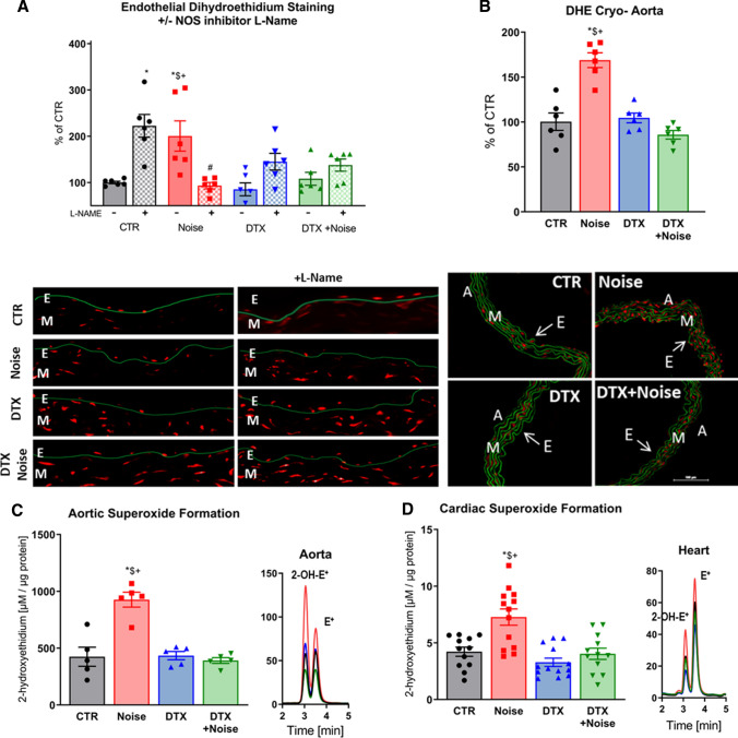
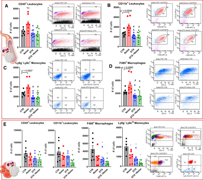
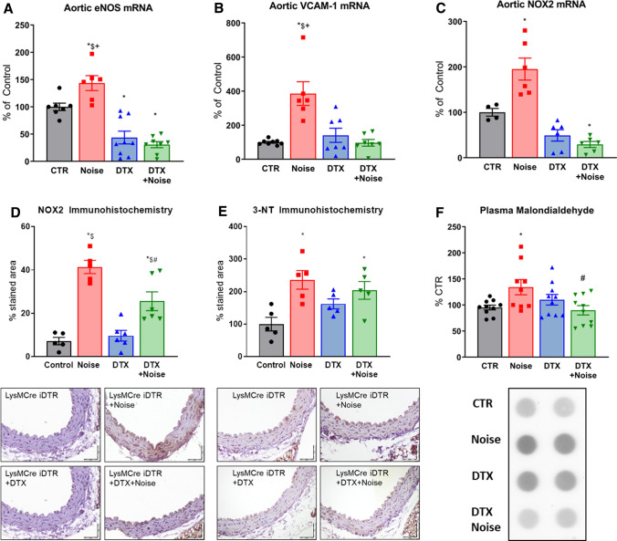
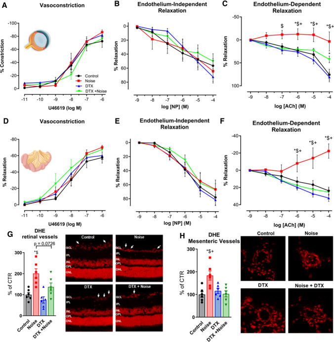
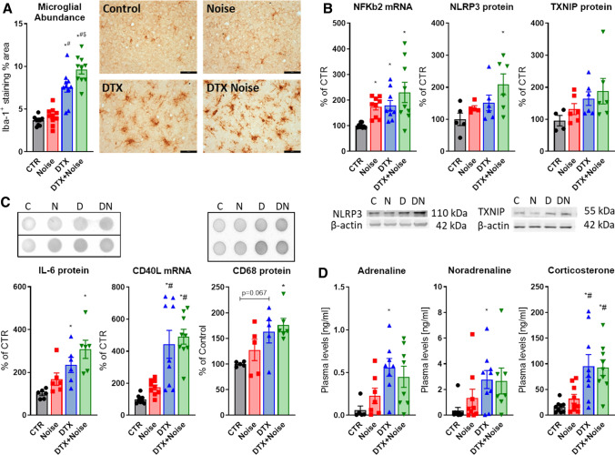
Aircraft noise induces vascular and cerebral inflammation and oxidative stress causing hypertension and cardiovascular/cerebral dysfunction. With the present studies, we sought to determine the role of myeloid cells in the vascular vs. cerebral consequences of exposure to aircraft noise. Toxin-mediated ablation of lysozyme M (LysM) myeloid cells was performed in LysMCre mice carrying a cre-inducible diphtheria toxin receptor. In the last 4d of toxin treatment, the animals were exposed to noise at maximum and mean sound pressure levels of 85 and 72 dB(A), respectively. Flow cytometry analysis revealed accumulation of CD45, CD11b, F4/80, and Ly6GLy6C cells in the aortas of noise-exposed mice, which was prevented by LysM cell ablation in the periphery, whereas brain infiltrates were even exacerbated upon ablation. Aircraft noise-induced increases in blood pressure and endothelial dysfunction of the aorta and retinal/mesenteric arterioles were almost completely normalized by ablation. Correspondingly, reactive oxygen species in the aorta, heart, and retinal/mesenteric vessels were attenuated in ablated noise-exposed mice, while microglial activation and abundance in the brain was greatly increased. Expression of phagocytic NADPH oxidase (NOX-2) and vascular cell adhesion molecule-1 (VCAM-1) mRNA in the aorta was reduced, while NFκB signaling appeared to be activated in the brain upon ablation. In sum, we show dissociation of cerebral and peripheral inflammatory reactions in response to aircraft noise after LysM cell ablation, wherein peripheral myeloid inflammatory cells represent a dominant part of the pathomechanism for noise stress-induced cardiovascular effects and their central nervous counterparts, microglia, as key mediators in stress responses.
飞机噪声可引起血管和大脑炎症以及氧化应激,导致高血压和心血管/大脑功能障碍。在本研究中,我们旨在确定髓样细胞在飞机噪声暴露导致血管和大脑后果中的作用。在携带 Cre 诱导白喉毒素受体的 LysMCre 小鼠中,用毒素介导的溶菌酶 M (LysM) 髓样细胞消融。在毒素治疗的最后 4 天,动物分别在最大和平均声压级为 85 和 72dB(A)的噪声下暴露。流式细胞术分析显示,在噪声暴露的小鼠中,CD45、CD11b、F4/80 和 Ly6G/Ly6C 细胞在主动脉中积累,这种积累在外周通过 LysM 细胞消融而被阻止,而大脑浸润在消融后甚至加剧。飞机噪声诱导的血压升高和主动脉、视网膜/肠系膜小动脉内皮功能障碍几乎完全通过消融得到纠正。相应地,在消融的噪声暴露小鼠中,主动脉、心脏和视网膜/肠系膜血管中的活性氧物质被减弱,而大脑中的小胶质细胞激活和丰度大大增加。主动脉、心脏和视网膜/肠系膜血管中吞噬 NADPH 氧化酶 (NOX-2) 和血管细胞黏附分子-1 (VCAM-1)mRNA 的表达减少,而 NFκB 信号似乎在消融后在大脑中被激活。总之,我们在 LysM 细胞消融后显示出对飞机噪声的中枢和外周炎症反应的分离,其中外周髓样炎症细胞代表了噪声应激诱导的心血管效应的发病机制的主要部分,而它们的中枢对应物,小胶质细胞,作为应激反应的关键介质。