Department of Agricultural and Biosystems Engineering, Iowa State University, Ames, IA, United States of America.
Department of Molecular Biology, Biochemistry and Biophysics, Iowa State University, Ames, IA, United States of America.
PLoS One. 2021 May 5;16(5):e0250650. doi: 10.1371/journal.pone.0250650. eCollection 2021.
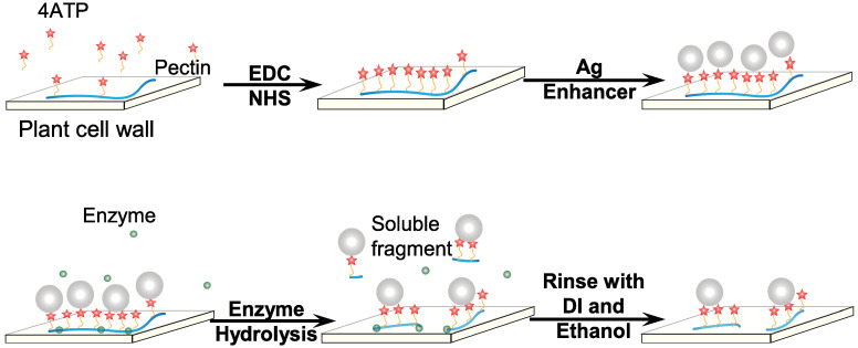
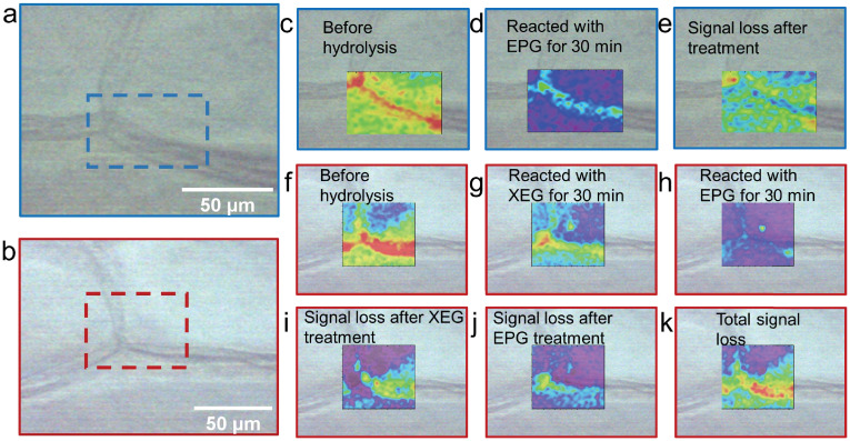
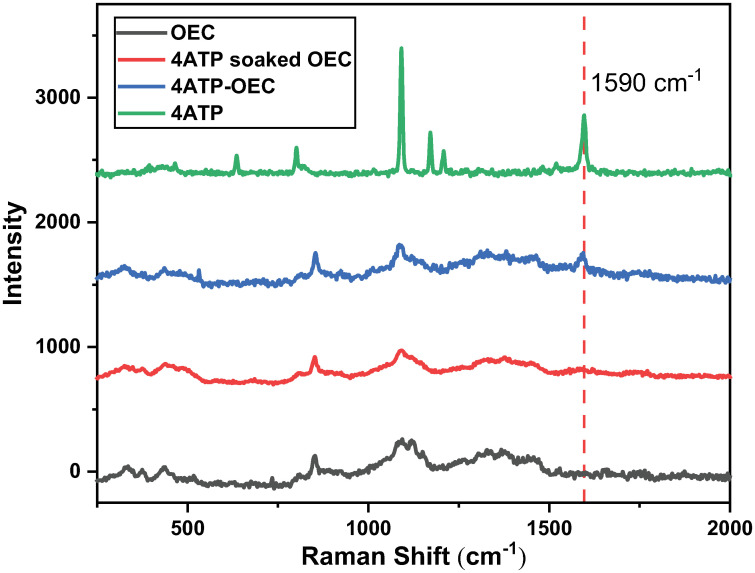
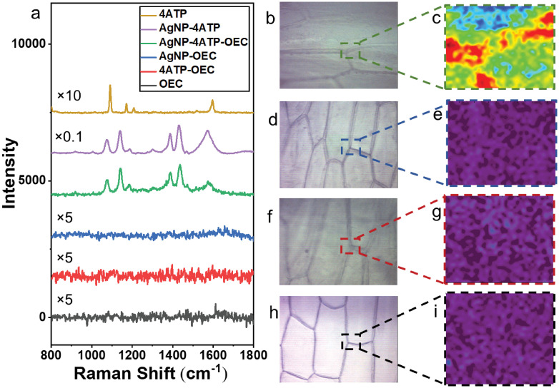
The primary plant cell wall is a complex matrix composed of interconnected polysaccharides including cellulose, hemicellulose, and pectin. Changes of this dynamic polysaccharide system play a critical role during plant cell development and differentiation. A better understanding of cell wall architectures can provide insight into the plant cell development. In this study, a Raman spectroscopic imaging approach was developed to visualize the distribution of plant cell wall polysaccharides. In this approach, Surface-enhanced Raman scattering (SERS through self-assembled silver nanoparticles) was combined with Raman labels (4-Aminothiophenol. 4ATP) and targeted enzymatic hydrolysis to improve the sensitivity, specificity, and throughput of the Raman imaging technique, and to reveal the distribution of pectin and its co-localization with xyloglucan inside onion epidermal cell (OEC) wall. This technique significantly decreased the required spectral acquisition time. The resulted Raman spectra showed a high Raman signal. The resulted Raman images successfully revealed and characterized the pectin distribution and its co-localization pattern with xyloglucan in OEC wall.
初生植物细胞壁是一个复杂的基质,由相互连接的多糖组成,包括纤维素、半纤维素和果胶。这个动态多糖系统的变化在植物细胞的发育和分化过程中起着关键作用。更好地了解细胞壁结构可以深入了解植物细胞的发育。在这项研究中,我们开发了一种基于拉曼光谱成像的方法,以可视化植物细胞壁多糖的分布。在该方法中,表面增强拉曼散射(通过自组装银纳米粒子实现的 SERS)与拉曼标记物(4-巯基苯胺,4-ATP)和靶向酶解相结合,以提高拉曼成像技术的灵敏度、特异性和通量,并揭示了在洋葱表皮细胞(OEC)壁内果胶的分布及其与木葡聚糖的共定位情况。该技术显著减少了所需的光谱采集时间。得到的拉曼光谱显示出很高的拉曼信号。所得的拉曼图像成功地揭示并表征了 OEC 壁中果胶的分布及其与木葡聚糖的共定位模式。