Department of Diagnostic Radiology & Nuclear Medicine, University of Maryland School of Medicine, Baltimore, MD, USA.
University of Liège, Belgium.
J Alzheimers Dis. 2021;81(4):1727-1735. doi: 10.3233/JAD-210116.
Cross-sectional studies have shown lower cerebral blood flow (CBF) in Alzheimer's disease (AD), but longitudinal CBF changes in AD are still unknown.
To reveal the longitudinal CBF changes in normal control (NC) and the AD continuum using arterial spin labeling perfusion magnetic resonance imaging (ASL MRI).
CBF was calculated from two longitudinal ASL scans acquired 2.22±1.43 years apart from 140 subjects from the Alzheimer's Disease Neuroimaging Initiative (ADNI). At the baseline scan, the cohort contained 41 NC, 74 mild cognitive impairment patients (MCI), and 25 AD patients. 21 NC converted into MCI and 17 MCI converted into AD at the follow-up. Longitudinal CBF changes were assessed using paired-t test for non-converters and converters separately at each voxel and in the meta-ROI. Age and sex were used as covariates.
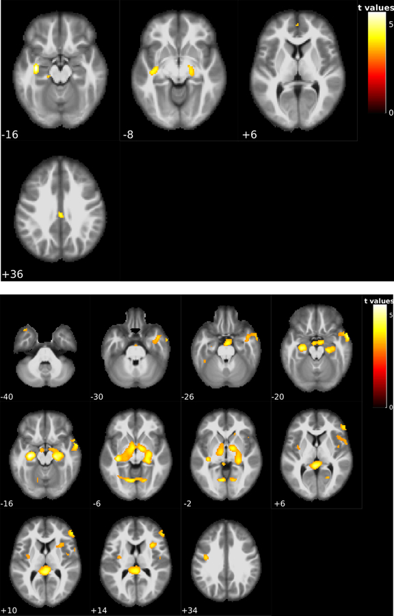
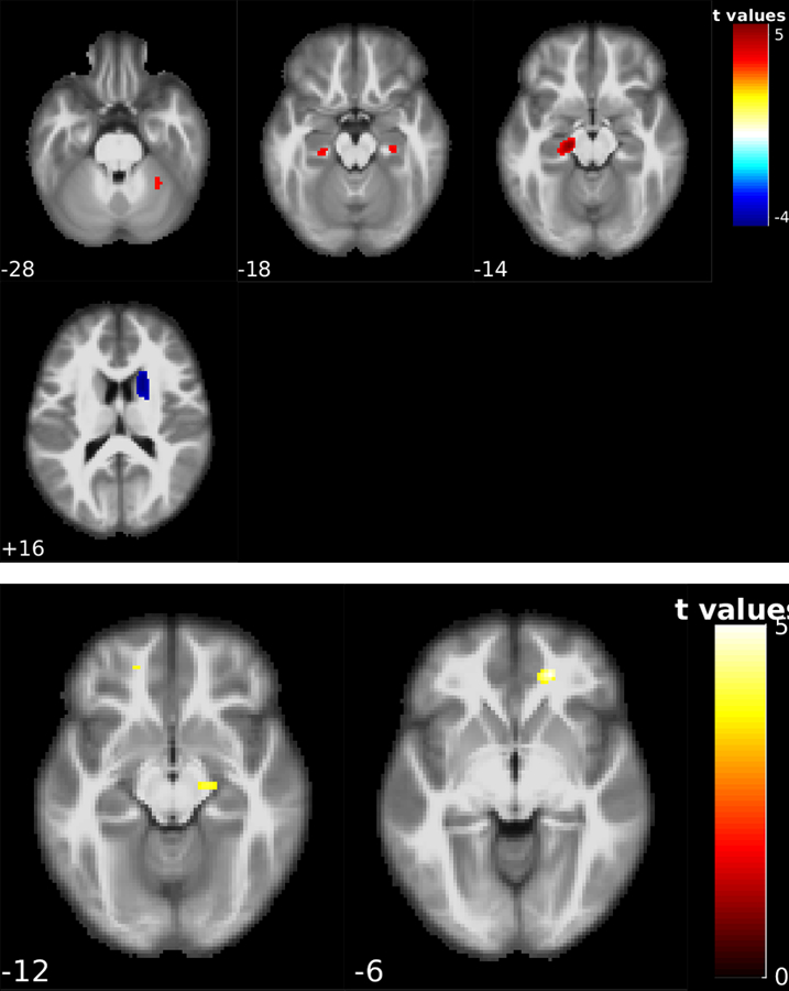
CBF reductions were observed in all subjects. Stable NC (n = 20) showed CBF reduction in the hippocampus and precuneus. Stable MCI patients (n = 57) showed spatially more extended CBF reduction patterns in hippocampus, middle temporal lobe, ventral striatum, prefrontal cortex, and cerebellum. NC-MCI converters showed CBF reduction in hippocampus and cerebellum and CBF increase in caudate. MCI-AD converters showed CBF reduction in hippocampus and prefrontal cortex. CBF changes were not related with longitudinal neurocognitive changes.
Normal aging and AD continuum showed common longitudinal CBF reductions in hippocampus independent of disease and its conversion. Disease conversion independent longitudinal CBF reductions escalated in MCI subjects.
横断面研究表明,阿尔茨海默病(AD)患者的大脑血流(CBF)较低,但 AD 的纵向 CBF 变化仍不清楚。
使用动脉自旋标记灌注磁共振成像(ASL MRI)揭示正常对照(NC)和 AD 连续体的纵向 CBF 变化。
从阿尔茨海默病神经影像学倡议(ADNI)的 140 名受试者中获取的两次纵向 ASL 扫描中计算 CBF,两次扫描的时间间隔为 2.22±1.43 年。在基线扫描时,队列包含 41 名 NC、74 名轻度认知障碍患者(MCI)和 25 名 AD 患者。21 名 NC 在随访中转化为 MCI,17 名 MCI 转化为 AD。分别在每个体素和meta-ROI 中使用配对 t 检验评估非转化者和转化者的纵向 CBF 变化,并使用年龄和性别作为协变量。
所有受试者均出现 CBF 降低。稳定的 NC(n=20)表现出海马体和后扣带回的 CBF 降低。稳定的 MCI 患者(n=57)表现出更广泛的空间分布的 CBF 降低模式,包括海马体、颞中回、腹侧纹状体、前额叶皮层和小脑。NC-MCI 转化者表现出海马体和小脑的 CBF 降低以及尾状核的 CBF 增加。MCI-AD 转化者表现出海马体和前额叶皮层的 CBF 降低。CBF 变化与纵向神经认知变化无关。
正常衰老和 AD 连续体在不依赖疾病及其转化的情况下,在海马体中表现出共同的纵向 CBF 降低。MCI 患者的疾病转化独立的纵向 CBF 降低呈上升趋势。