Shah Sonalee, Mishra Biswajit, Tiwari Nidhi, Nikunj Anand
Department of Oral Pathology, Government Dental College, Raipur, Chhattisgarh, India.
Department of Oral Surgery, Government Dental College, Raipur, Chhattisgarh, India.
J Oral Maxillofac Pathol. 2020 Sep-Dec;24(3):589. doi: 10.4103/jomfp.JOMFP_142_20. Epub 2021 Jan 9.
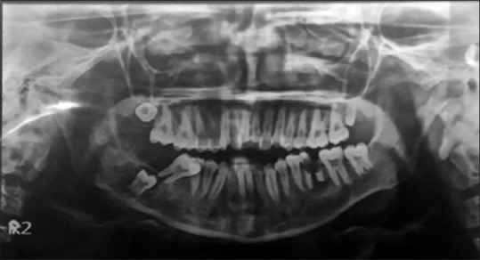
Osteosarcoma (OS) accounts for about 20% of all sarcomas with gnathic involvement seen in about 6%-10% of all OSs. The clinical presentation of OSs in the jaws is different from that of long bones as swelling is the most common complaint in patients with jaw OS followed by pain. The histopathologic variables seen are more favorable in OSs of jaws. Low-grade tumors are Stage I, high-grade tumors are Stage II and metastatic tumors (regardless of grade) are Stage III. A 17-year-old male patient reported with a complaint of the presence of an intra-oral growth gradually increasing in size in the right buccal mucosa region soft tissue enveloping the occlusal aspect of the right mandibular second molar. Extraorally swelling was present on the right side of the face for 4 months. Radiographically, there was a radiolucency from the distal aspect of right Mandibular second molar extending into the ramus region of the mandible with ill-defined borders. Hemi-mandibulectomy was done with the removal of the right mandible from the premolar region to condyle and coronoid processes. Microscopic evaluation of the sections after hematoxylin and eosin staining revealed interlacing fascicles of spindle-shaped cells arranged in a biphasic pattern and some areas of attempted bone formation evident in deeper sections. Tumor was an osteoblastic variety consisting of tumor osteoid surrounded by bizarrely arranged fibroblast-like cells. It showed positive staining with α-smooth muscle actin and Vimentin, suggesting a malignant tumor of mesenchymal cells with high myofibroblastic activity. Our case had small-cell histology; therefore, differential diagnosis was important.
骨肉瘤(OS)约占所有累及颌骨的肉瘤的20%,在所有骨肉瘤中,约6%-10%会累及颌骨。颌骨骨肉瘤的临床表现与长骨骨肉瘤不同,肿胀是颌骨骨肉瘤患者最常见的主诉,其次是疼痛。颌骨骨肉瘤的组织病理学变量更有利。低级别肿瘤为I期,高级别肿瘤为II期,转移性肿瘤(无论级别)为III期。一名17岁男性患者主诉右侧颊黏膜区域软组织内有一口腔内肿物,其大小逐渐增大,该软组织包绕右侧下颌第二磨牙的咬合面。右侧面部口外肿胀已有4个月。影像学检查显示,右侧下颌第二磨牙远中侧有一透亮区,延伸至下颌升支区域,边界不清。行半侧下颌骨切除术,从第一前磨牙区域至髁突和喙突切除右侧下颌骨。苏木精和伊红染色后对切片进行显微镜评估,可见梭形细胞交织成束,呈双相模式排列,在更深的切片中可见一些试图形成骨的区域。肿瘤为成骨细胞型,由肿瘤类骨质组成,周围有排列怪异的成纤维细胞样细胞。它对α-平滑肌肌动蛋白和波形蛋白呈阳性染色,提示为具有高肌成纤维细胞活性的间充质细胞恶性肿瘤。我们的病例为小细胞组织学;因此,鉴别诊断很重要。